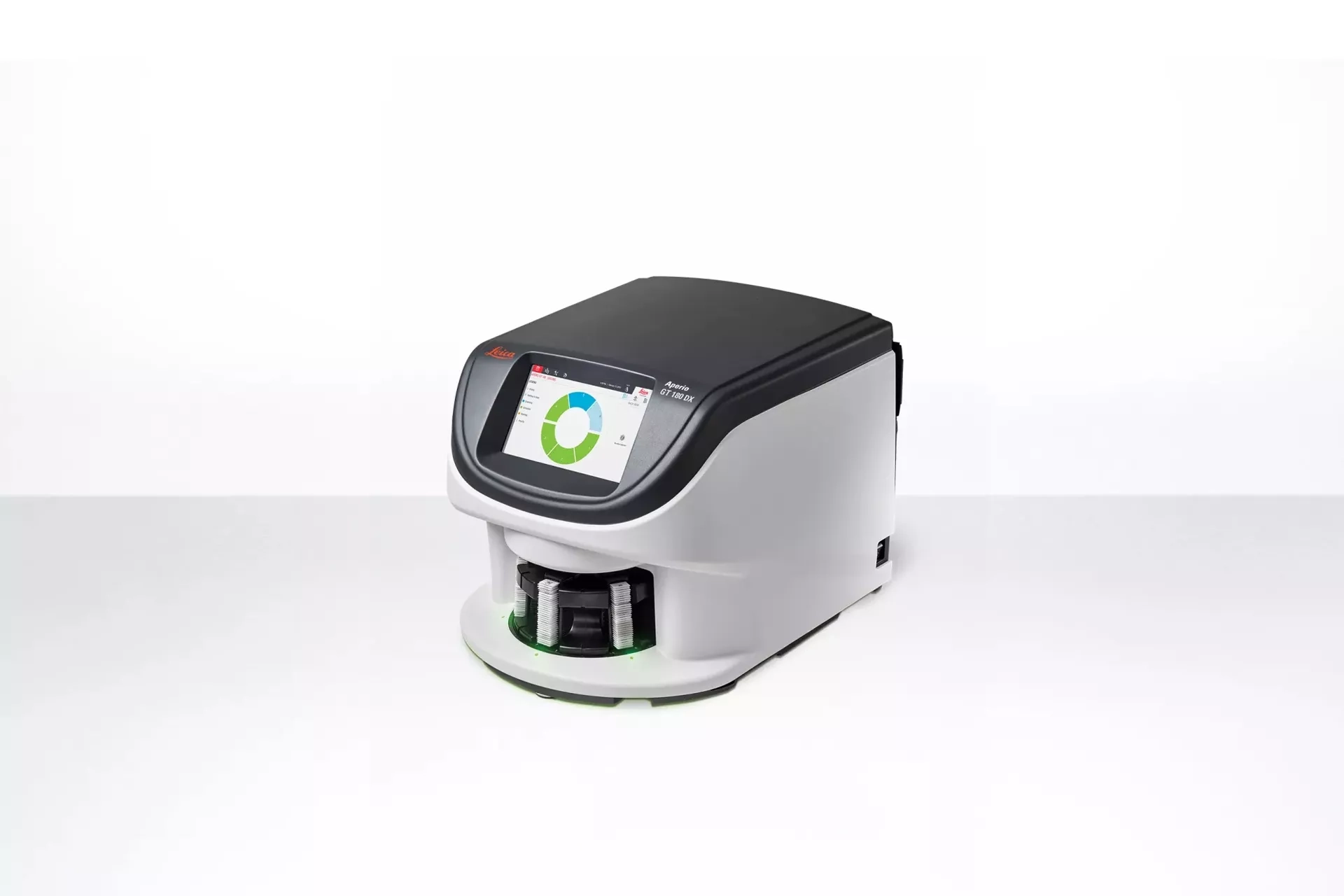
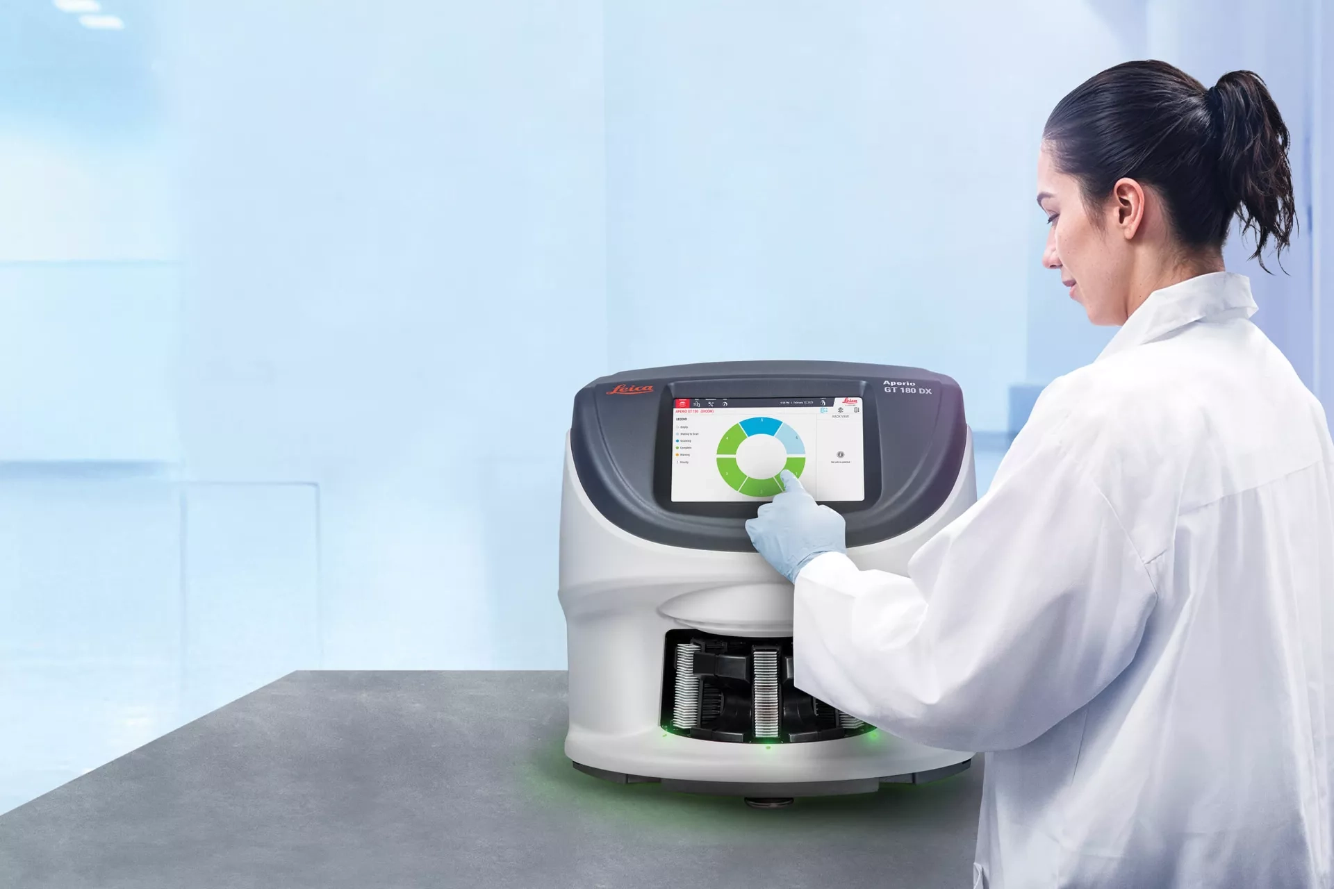
Leica Biosystems – Aperio GT 180 DX Scanner เครื่องสแกนสไลด์ชิ้นเนื้อ สำหรับงานพยาธิวิทยา
ถึงเวลาพลิกมุมมอง 180 องศา เพราะเทคโนโลยีที่ผ่านการพิสูจน์แล้ว นำไปสู่พยาธิวิทยาที่ดียิ่งกว่า
Aperio GT 180 DX มอบคุณภาพของภาพและความเร็วในการสแกนที่เป็นเอกลักษณ์ของเครื่องสแกนในตระกูล Aperio ที่ได้รับความไว้วางใจทั่วโลก ในรูปแบบการออกแบบที่เหมาะสมกับห้องปฏิบัติการพยาธิวิทยาคลินิกสมัยใหม่
สำหรับนักพยาธิวิทยาหลายท่าน การเปลี่ยนผ่านสู่ Digital Pathology อาจยังคงเป็นเรื่องท้าทาย ท่ามกลางแรงกดดันที่เพิ่มขึ้นในการทำงานให้ได้มากขึ้นด้วยทรัพยากรที่จำกัด พร้อมทั้งต้องรักษาความรวดเร็ว ความสม่ำเสมอ และความสามารถในการทำงานร่วมกัน
ห้องปฏิบัติการยุคใหม่จึงต้องการเครื่องสแกนสไลด์ดิจิทัลที่มีขนาดกะทัดรัด คุ้มค่า และสามารถให้ผลลัพธ์คุณภาพสูงได้โดยไม่จำเป็นต้องอาศัยการฝึกอบรมในระดับผู้เชี่ยวชาญ
คุณสมบัติและประโยชน์เด่นของ Aperio GT 180 DX
- ใช้งานง่าย (Easy-to-Use)
เรียนรู้การใช้งานได้รวดเร็ว ใช้เวลาในการฝึกอบรมน้อย - การออกแบบแบบกะทัดรัดและครบวงจร (Compact, Integrated Design)
มาพร้อมหน้าจอสัมผัสในตัว ไม่จำเป็นต้องใช้คอมพิวเตอร์หรือจอภาพภายนอกเพิ่มเติม - รองรับการโหลดสไลด์ระดับกลาง (Mid-Capacity Loading)
รองรับการสแกนได้สูงสุด 180 สไลด์ พร้อมระบบเติมแร็กสไลด์ได้อย่างต่อเนื่อง - คุณภาพทางออปติกระดับเยี่ยม (Excellent Optical Quality)
ด้วยเลนส์คุณภาพสูงจาก Leica ให้ภาพ Whole Slide Image ที่คมชัดและมีความละเอียดสูง - รองรับ DICOM และ Z-Stack*
รองรับการสแกนที่กำลังขยาย 20x และ 40x พร้อมการแสดงผลหลายชั้นโฟกัส เพื่อการวิเคราะห์รายละเอียดที่แม่นยำยิ่งขึ้น - Manual Scan และ Extended Focus*
เครื่องมือเสริมสำหรับการปรับแต่งคุณภาพภาพ การสแกนซ้ำ และการเพิ่มความคมชัดในตัวอย่างที่มีความท้าทาย - สแกนได้อย่างรวดเร็ว (Fast Scanning)
สแกนพื้นที่ขนาด 15 x 15 มม. ที่กำลังขยาย 40x ได้ภายในเวลาเพียง 32 วินาที - ระบบควบคุมคุณภาพด้วย AI (AI Quality Control)
ซอฟต์แวร์ Aperio iQC Software* ใช้ปัญญาประดิษฐ์ในการตรวจจับและแจ้งเตือนปัญหาด้านคุณภาพของภาพโดยอัตโนมัติ
It’s Time for a 180 – Because Proven Technology Means Better Pathology
The Aperio GT 180 DX scanner delivers the excellent image quality and speed of trusted Aperio scanners in a design that works for your clinical laboratory.
For many pathologists, the shift to digital pathology still feels like a daunting task. Teams are navigating increasing pressure to do more with less, all while balancing tight timelines and growing expectations around collaboration and consistency. Modern labs need a digital pathology slide scanner that’s compact, cost-effective, and capable of delivering high-quality results without expert-level training.
Key Features and Benefits of the Aperio GT 180 DX Scanner
- Easy-to-Use – limited training required.
- Compact, Integrated Design – Built-in touchscreen; no external PC or monitor required.
- Mid-Capacity Loading – Scans up to 180 slides with continuous rack access.
- Excellent Optical Quality – Leica optics deliver sharp, high-quality whole slide images.
- DICOM & Z-Stack Support* – 20x/40x scanning and multi-layer viewing.
- Manual Scan & Extended Focus* – Optional tools for image clarity and fine adjustments.
- Fast Scanning – Scans a 15mm x 15mm area in 32 seconds at 40x.
- AI Quality Control – Aperio iQC Software* detects and flags image issues.

Manual Slide Scanning*
Gain control over slide capture, which helps ensure high-quality images from even the most challenging tissue samples. Quickly rescan slides from the same location, making real-time adjustments to faint stains, small tissue fragments, or low-density samples. This feature minimizes workflow disruptions and enhances image clarity and consistency.
Aperio iQC DX Software*
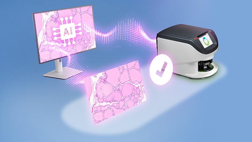
Optional Aperio iQC DX software enhances automatic image quality control, ensuring confidence before scans are delivered. Using AI-enabled detection, it identifies whole slide image (WSI) artifacts and provides notifications through a quality control dashboard, flagging issues and recommending corrective actions so anyone in the lab can rescan.
Extended Focus*
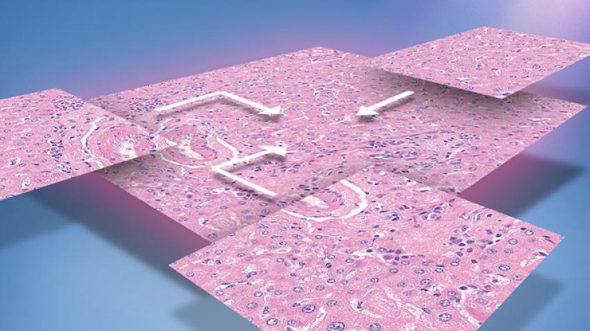
An efficient imaging solution that enhances image quality while optimizing storage needs. By combining multiple focus layers into a single, ultra-sharp image, Extended Focus ensures all cells remain in focus without requiring users to scroll through layers. Smaller file sizes speed up data transfer, facilitating faster collaboration and enabling remote access to high-quality images.
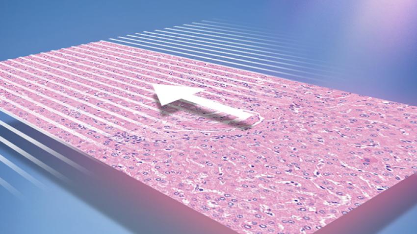
Default Calibration Point
Default Calibration Point reduces the risk of striping from the start, allowing lab managers to maintain image clarity without additional adjustments. technicians benefit from a streamlined scanning process that enhances efficiency and delivers consistently high-quality results with minimal effort.
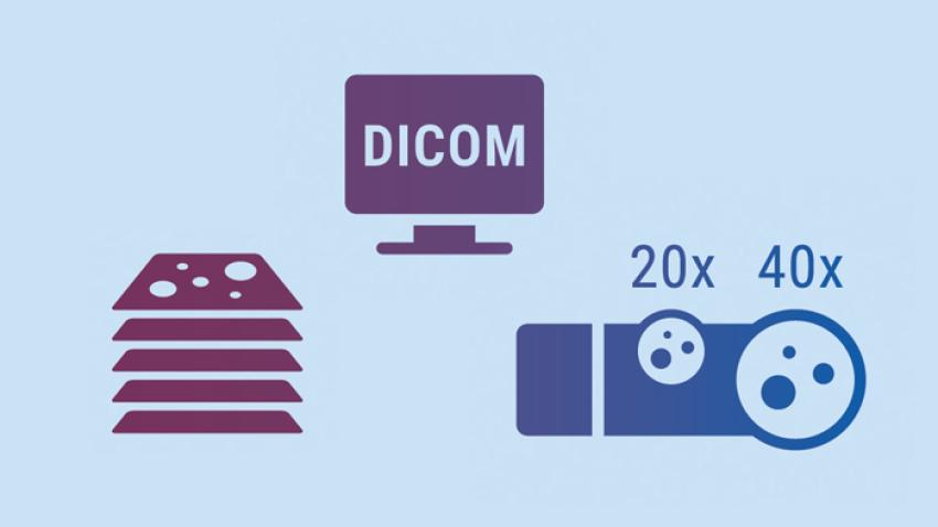
DICOM with 20x,40x and Z-Stack*
Aperio GT 180 scanner uses a high-performance objective manufactured by Leica Microsystems, which has produced world-class optics since 1847. Leica optics deliver exceptional image quality.
- Available alongside .svs, DICOM enables image clarity, improved collaboration, and a streamlined workflow.
- 20x and 40x options for clear and detailed views of samples
- Z-Stacking enables the review of slide samples at varied heights
*Optional Features
It’s Time for a 180 Because Proven Technology Means Better Pathology
Twenty-five+ years of digital pathology expertise can help everyone in your clinical lab take the next step into digital pathology with confidence. With more than 70 third-party software integrations, 9 generations of scanning pedigree, and 380+ patents, our innovation ensures interoperability and efficiency across your entire pathology workflow.
For Pathologists
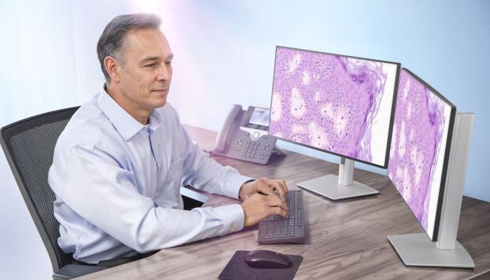
Deliver diagnostic confidence with expertly engineered Leica optics, producing consistently high-quality digital slides that reveal critical details, even in complex cases. Seamlessly integrated into clinical workflows, the Aperio GT 180 DX scanner enables rapid case turnaround, streamlined collaboration, and reproducible results that support timely, informed diagnoses.
For Lab Managers
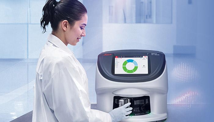
Easy to use and intuitive to learn, the continuous loading, high-automation, and small footprint enables labs to enjoy a smooth, stress-free digital pathology workflow. Advanced feature options such as Manual Scanning*, for challenging slides, and Aperio iQC DX software*, to streamline quality control review, ensure hassle-free, high-quality scans.
For IT Managers
Secure, scalable, and easy to integrate; seamlessly connect the Aperio GT 180 DX scanner to your lab’s existing systems and infrastructure and leverage the widely used .svs or standardized DICOM file format for rapid integration with downstream software.
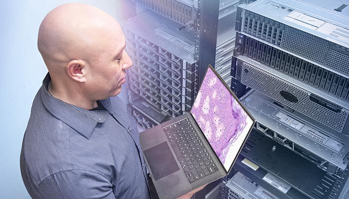
Unrivaled Whole Slide Image Quality
Capture every critical detail with exceptional clarity, from cellular grooves and chromatin structure to intracellular bridges in H&E, or crisp, clear images of your IHC, ISH, and brightfield multiplex slides.
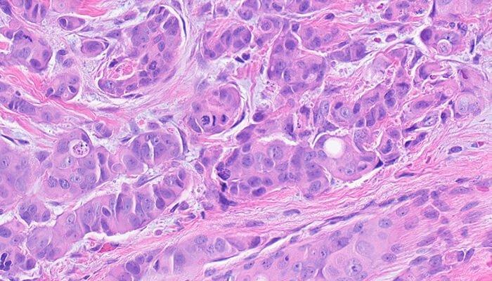
Mitotic Events (Breast)
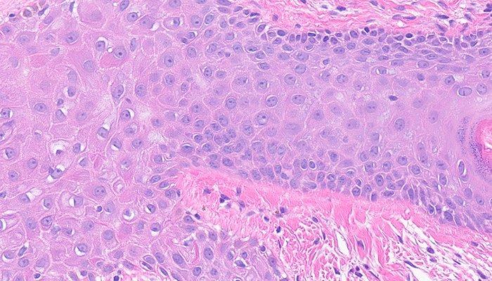
Intercellular Bridges
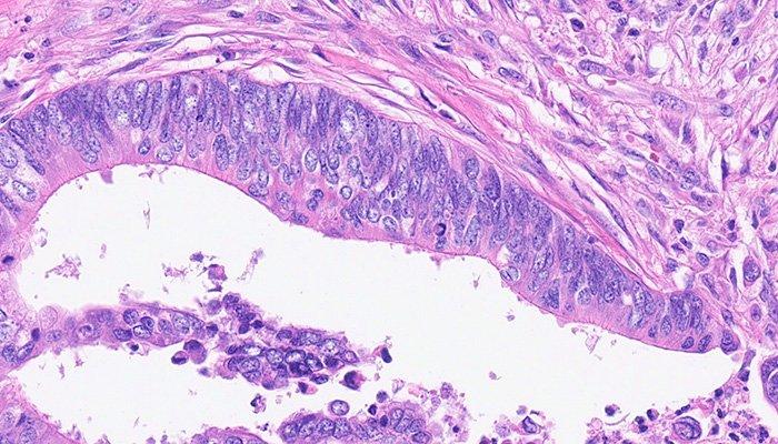
Chromatin
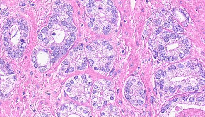
Nucleoli
Precision, Potential to Scale, and Peace of Mind
Digital pathology scan speeds have not always been fast enough at 40x magnification to keep up with high-volume scanning (120k+ slides per year).
Real-Time Focusing (RTF)** offers a potential solution to this problem. It’s a novel method to capture a digital image that combines an imaging line sensor and a focusing line sensor.
- While scanning, the focusing line sensor receives focusing data of the tissue.
- Proprietary algorithms determine the best focus value.
- Then, each best focus value is treated like a control loop with the mechanics, feeding the best focus value to the objective height on the fly.
- The positioning adjustments of the objective are performed continuously (in real time) in order to enable the imaging line sensor to capture the image at its best possible focus. Hence, real time focusing.
**US Patent no. 9,841,590