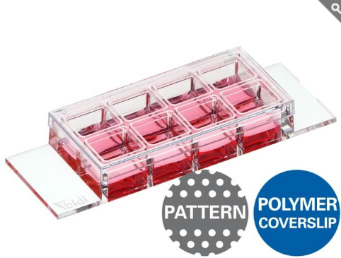
คุณสมบัติเด่น
- พื้นผิวไมโครแพทเทิร์น (Micropatterned Surface) สำหรับการศึกษาพฤติกรรมเซลล์ในรูปแบบ 1 มิติ (1D Cell Assays) และการถ่ายภาพความละเอียดสูง
- ออกแบบลวดลายไมโครสเกลแบบเส้นตรง (Line Micropatterning) เพื่อจำกัดพื้นที่การยึดเกาะของเซลล์ให้อยู่ในแนวหนึ่งมิติ ช่วยให้สามารถศึกษาการเคลื่อนที่ รูปร่าง และการจัดเรียงตัวของเซลล์ได้อย่างแม่นยำ
- ปรับให้เหมาะสมสำหรับงานกล้องจุลทรรศน์ ด้วยคุณสมบัติทางแสงที่ยอดเยี่ยม ให้ภาพคมชัดและมีความละเอียดสูง
- พร้อมใช้งานทันที (Ready-to-Use) และผ่านการทำให้ปราศจากเชื้อ (Sterile) ช่วยลดขั้นตอนการเตรียมตัวอย่าง เพียงแกะบรรจุภัณฑ์และเริ่มการทดลองได้ทันที
Micropatterned surface for 1D cell assays and high-resolution imaging
- Line micropatterning with a confined one-dimensional adhesion area
- Optimized for microscopy with exceptional optical clarity
- Ready-to-use & sterile: Unpack-and-start
Applications
- Defined One-Dimensional (1D) Cell Positioning on Microlanes – Enables precise spatial confinement and imaging of single cells or cell clusters
- Single-Cell Polarization and Migration Assays – Ideal for studying geometry-driven migration, cytoskeletal dynamics, and organelle trafficking
- Neuronal Cell and Organelle Outgrowth – Physical boundaries support applications such as neurite extension, neuronal projections, and cell–cell communication.
- Cell Cluster Polarization – Facilitates the orientation and interaction of epithelial and endothelial cells along defined adhesive patterns
- Precise Cell Alignment – Ideal for microscopy-based transfection studies, proteomics, and metabolic activity assays
- Live Cell and Fluorescence Imaging – Optimized for high-resolution long-term or fluorescence microscopy of both living and fixed cells
- Immunofluorescence stainings – Facilitates detailed protein localization and cell signaling studies
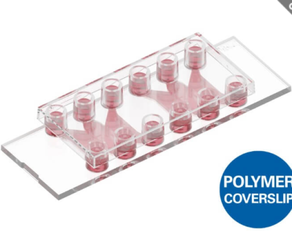
- Labware Format: also available in the µ-Slide VI 0.4 Channel-Slide format – suitable for flow and shear stress applications (#83614)
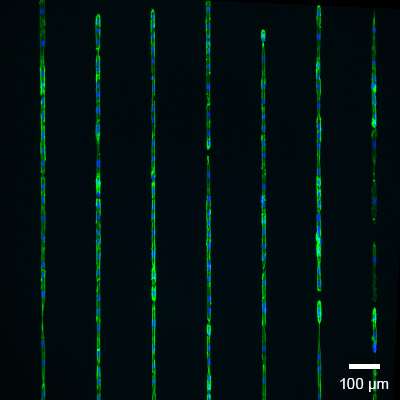
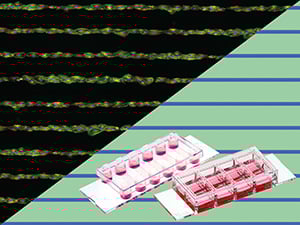
Human Umbilical Vein Endothelial Cells (HUVEC) on the 20 µm lines. Pre-coating with Matrigel® before cell seeding. After an incubation period of two days, fixation and staining were applied. Blue: Cell nuclei (DAPI), green: F-actin cytoskeleton (Alexa 488 conjugate), objective lens 10x, widefield fluorescence.
Good to Know!
ibiTreat patterns can be used directly or coated with extracellular matrix proteins, such as laminin!
The application of specific protein coatings (e.g., laminin) on ibiTreat is straightforward and comparable to the process used for our non-patterned ibiTreat µ-Slides.
Specifications
| µ-Slide geometry | See product page µ-Slide 8 Well high |
| Binding motif | ibiTreat (TC-treated) |
| Pattern shape | Line |
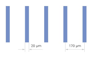
| Line width | 20 µm |
| Pitch | 170 µm |
| Pattern layout | perpendicular to the slide length |
| Surface passivation | Bioinert |
| Number of patterns | ca. 63 per well |
| Bottom material | #1.5 ibidi Polymer Coverslip |
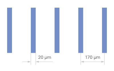
Technical Features
- Precision µ-Patterning – The µ-Slide features an ibiTreat micropatterned surface enclosed by an advanced ibidi Bioinert surface, ensuring selective cell adhesion.
- Exclusive Bioinert Surface – Outperforms standard ultra-low attachment (ULA) surfaces:
- Ensures an environment free from any unwanted adhesion of cells or proteins
- Long-term stability for days or weeks
- Biologically inert, preserving cell viability and behavior
- Superior Imaging Compatibility – The Bioinert surface is applied to the ibidi Polymer Coverslip, delivering unmatched optical clarity for high-resolution microscopy.
- Non-Fluorescent, Phase Contrast Compatible – Patterns remain non-fluorescent but slightly visible under phase contrast, facilitating easy alignment and analysis.
- Compatibility – Fully biocompatible materials, compatible with different protein and peptide coatings as well as with staining and fixation solutions
- Different Slide Formats – Available on either the µ-Slide 8 Well high or µ-Slide VI 0.4

Interested in unlocking new possibilities with ibidi µ-Patterning?
Let’s collaborate on a custom solution to meet your research needs! We will work with you to co-develop a tailor-made design here.
You need support or want to test the ibiTreat µ-Patterns first? Contact us or choose up to three free samples from our ibiTreat micropatterning standard products!
The Principle of the µ-Patterning Technology
The ibidi µ-Patterning technology provides spatially defined cell adhesion for various spheroid and organoid applications. The miniaturized, adhesive patterns are irreversibly printed in the non-adhesive Bioinert surface of the ibidi Polymer Coverslip, which allows for precisely controlled cell adhesion. The µ-Patterns are dry-stable, sterile, and ready to use. The ibiTreat µ-patterns are slightly visible under the phase contrast microscope but not visible using fluorescence microscopy.
ibiTreat is the hydrophilic, tissue culture-treated (TC-treated) version of the ibidi Polymer Coverslip. This modification of the physical surface, similar to the treatment used in standard cell culture vessels, creates a hydrophilic surface and ensures adherence to nearly all cell types.
The application of specific protein coatings (e.g., laminin) on ibiTreat is straightforward and comparable to the process used for our non-patterned ibiTreat µ-Slides.
The µ-Pattern, the Bioinert surface, and the ibidi Polymer Coverslip are all optimized for high-resolution imaging and microscopy.
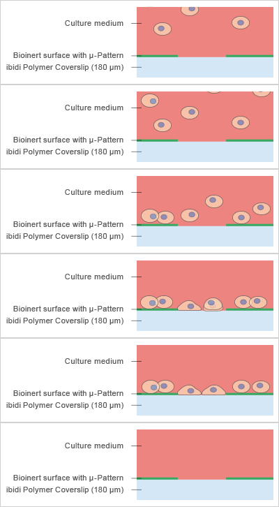
Applications of Line µ-Patterning
Micropatterns are powerful tools for creating one-dimensional (1D) cell assays, enabling precise control over cell localization and polarization. This approach opens new possibilities for studying cellular behavior in a defined 1D microenvironment. The combination of the flat-bottom design of ibidi’s µ-Slides and Bioinert surface passivation ensures exceptional optical clarity, making it ideal for high-resolution fluorescence microscopy.
Using ibidi µ-Patterns technology, linear patterns can be created from a cell suspension, directing cells to adhere and grow specifically along the 1D microlanes.
Application Examples
Human Umbilical Vein Endothelial Cells (HUVEC) on the 20 µm lines. Pre-coating with Matrigel ® before cell seeding. After an incubation period of two days, fixation and staining were applied. Blue: Cell nuclei (DAPI), green: F-actin cytoskeleton (Alexa 488 conjugate), objective lens 10x, widefield fluorescence.
HT-1080 cells grown on the μ-Slide 8 Well high μ-Pattern ibiTreat, lin20, pit170, precoated with Laminin. Cells exhibit guided migration constrained to the micropatterned lines, maintaining alignment and directionality without deviating from the defined tracks.
The ibiTreat µ-Pattern Variety
Understanding the Key Elements of µ-Patterns
Every ibidi µ-Pattern is designed with precise spatial control, ensuring reproducible cell adhesion and organization. Let’s break down the essential pattern components:
- Shape – Defines the geometry of the attachment area (e.g., cir = circle)
- Size – The diameter or width of the attachment area, measured in micrometers (µm) (e.g., 500 µm)
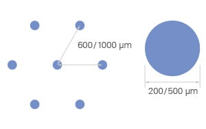
- Pitch – The spacing between adjacent shapes, measured from center to center (e.g., pit1000 = 1000 µm)
- Layout – The overall pattern arrangement, determining how attachment areas are structured (e.g., a hexagonal layout)
- Surface – The cell attachment surface where cells adhere, made of ibiTreat for optimal cell growth
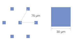
Single-Cell Arrays
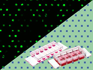
Square area
sqr30, pit75
Multi-Cell Arrays
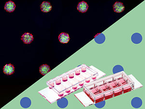
Circle area
cir200/500, pit600/1000
Line Arrays
Lines
lin20, pit170
Looking for other pattern options? Make a special request here, and we will develop a solution tailored to your specific applications.
Packaging Information
| Order No. | Packaging Unit | Packaging Dimensions (l × w × h) | Shipping Weight |
| 83814 | 10 (indiv. packed) | 150 × 130 × 110 mm | 0.2 kg |