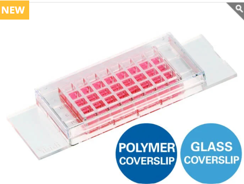
- แชมเบอร์พร้อมแผ่น Coverslip แบบ 32 หลุม สำหรับการเพาะเลี้ยงเซลล์ การย้อมอิมมูโนฟลูออเรสเซนซ์ (IF) และการถ่ายภาพด้วยกล้องจุลทรรศน์ความละเอียดสูง
- สไลด์สำหรับงานกล้องจุลทรรศน์ที่มี 32 หลุม จัดวางในรูปแบบเดียวกับ 384-well plate ช่วยให้เปลี่ยนจากการทดลองขนาดเล็กไปสู่การทดสอบแบบ High-Throughput ได้อย่างสะดวก
- พื้นที่เพาะเลี้ยงขนาดเล็ก ใช้จำนวนเซลล์และปริมาตรรีเอเจนต์น้อย ช่วยลดต้นทุนการทดลอง
- เหมาะอย่างยิ่งสำหรับการย้อม Immunofluorescence (IF) ได้อย่างง่ายดาย ด้วยแผ่น coverslip ที่รวมอยู่ในตัวผลิตภัณฑ์ ช่วยให้ได้ภาพคุณภาพสูงสำหรับการวิเคราะห์ด้วยกล้องจุลทรรศน์ความละเอียดสูง
- จุดเด่น
- รองรับงานเพาะเลี้ยงเซลล์และการวิเคราะห์ด้วยฟลูออเรสเซนซ์
- ประหยัดเซลล์และสารเคมี
- ให้คุณภาพภาพยอดเยี่ยมสำหรับการถ่ายภาพระดับเซลล์
- รองรับการขยายงานวิจัยไปสู่การคัดกรองตัวอย่างจำนวนมาก (High-Throughput Screening) ได้อย่างมีประสิทธิภาพ
Chambered coverslip with 32 wells for cell culture, IF, and high-resolution microscopy
- Microscopy slide with 32 wells in a 384-well plate geometry for easy transition to high-throughput
- Small footprint for low cell numbers and minimal reagent volumes
- Ideal for easy immunofluorescence staining with an integrated coverslip
Small-Volume Assay Development in a Microscopy Slide Format
The µ-Slide 32 Well is a chambered coverslip for developing, optimizing, and imaging 32 parallel cell-based assays on a single microscopy slide. Multiple experimental conditions, concentrations, staining parameters, and biological replicates can be tested while using only small numbers of cells and low reagent volumes.
The 32 wells are arranged according to the geometry of a standard 384 well format. This makes the µ-Slide 32 Well particularly useful for assay development and optimization before scaling up to high-throughput workflows.
Seamless Transition to Multiwell Formats
Develop and optimize cell-based assays in a convenient microscopy slide format. The well geometry and volume facilitate the transfer of established protocols to 384 well microplates (e.g., the ibidi µ-Plate 384 Well or µ-Plate 384 Well Clear) for automated high-content and high-throughput workflows.
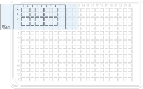
The wells of the µ-Slide 32 Well match the size and spacing of a standard 384 well plate, enabling easy transfer of cell models, protocols, and results from slide-based experiments to automated plate-based workflows.
High-Resolution Microscopy of Immunofluorescence Staining
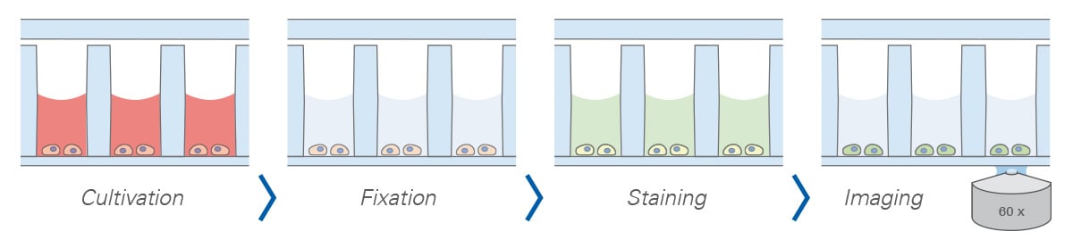
The ibidi µ-Slide 32 Well allows standard immunofluorescence protocols to be performed directly in the slide format. All steps: cell cultivation, fixation, staining, and imaging occur in the open well geometry. After staining, samples are imaged through the coverslip bottom using high-resolution microscopy.
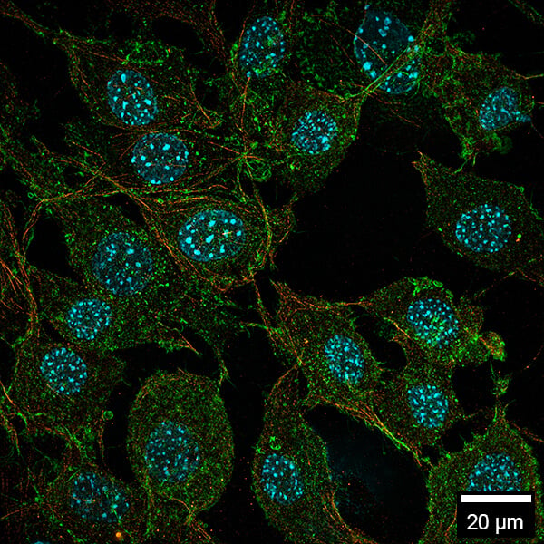
MCF7 cells in the µ-Slide 32 Well Glass Bottom, acquired using a ZEISS Elyra 7 microscope. Images were recorded sequentially with Nyquist sampling in SIM Apotome mode, followed by SIM2 processing, and displayed as a maximum intensity projection. Cell nuclei were stained with DAPI (blue), actin with phalloidin (green), and microtubules with a monoclonal antibody (red). 40× objective lens. Scale bar: 20 µm.
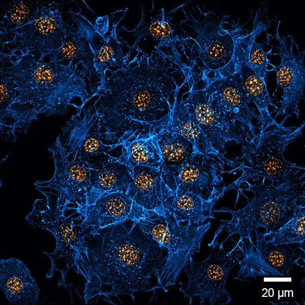
NIH-3T3 cells in the µ-Slide 32 Well ibiTreat, acquired using a ZEISS Elyra 7 microscope. Images were recorded sequentially with Nyquist sampling in SIM Apotome mode, followed by SIM2 processing, and displayed as a maximum intensity projection. Cell nuclei were stained with DAPI and shown in orange; F-actin was stained with phalloidin and shown in blue. 20× objective lens. Scale bar: 20 µm.
Complete Imaging Workflow: All in One Slide
High-Content Screening in a Slide Format
The µ-Slide 32 Well combines parallelization with a large accessible imaging area, making it well suited for imaging-based assay development and high-content analysis. Using a 63× objective and a 100 µm × 100 µm field of view, up to 1225 fields of view can be acquired per well.
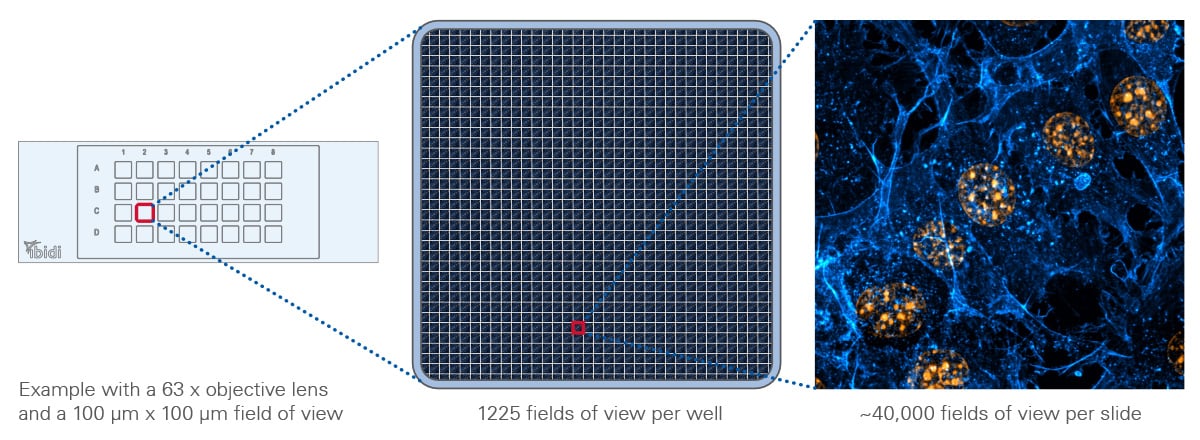
The µ-Slide 32 Well supports high-content imaging with up to 1225 fields of view per well, corresponding to approximately 40,000 fields of view across the entire slide.
Cell Culture and Advanced Microscopy
Cells can be cultured and treated across 32 independent experimental conditions on a single microscopy slide for applications such as immunofluorescence imaging, transfection assays, compound treatments, antibody optimization, and the comparison of biological replicates. Because the cells can remain in the same wells from cultivation over treatment, they can directly be imaged through the #1.5 ibidi Polymer Coverslip or #1.5H Glass Coverslip Bottom using brightfield microscopy, widefield fluorescence microscopy, confocal microscopy and many more. For techniques with particularly demanding optical requirements, such as TIRF and super-resolution microscopy, the #1.5H Glass Coverslip variant is recommended.
Specifications
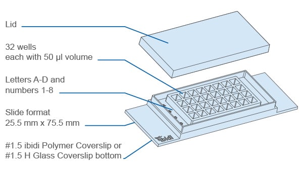
| Outer dimensions (w × l) | 25.5 × 75.5 mm |
| Number of wells | 32 |
| Dimensions of wells (w × l × h) | 3.5 × 3.5 × 6.8 mm |
| Volume per well | 50 µl |
| Height with/without lid | 8.2 / 7.0 mm |
| Growth area per well | 0.12 cm² |
| Coating area per well using 50 µl | 0.69 cm² |
| Bottom material | #1.5 ibidi Polymer Coverslip (different surfaces available) #1.5H Glass Coverslip (uncoated and untreated) |
For technical drawings and details, please see:
Instructions µ-Slide 32 Well (PDF)
Instructions µ-Slide 32 Well Glass Bottom (PDF)
Technical Features
Precision, Quality, and Biocompatibility
- µ-Slide (chambered coverslip) with 32 independent wells
- #1.5 ibidi Polymer Coverslip or #1.5H Glass Coverslip bottom with excellent optical quality
- Individual well walls minimize well-to-well crosstalk and contamination
- Wells are sized and distributed like on a 384 Well plate, e.g., the ibidi µ-Plate 384 Well or µ-Plate 384 Well Clear
- Compatible with multichannel pipettes
- Compatible with staining and fixation solutions
- Compatible with immersion oil
- Made from non-cytotoxic, biocompatible materials
- Sterile with single packaging
Product Versions
- With #1.5 ibidi Polymer Coverslip (different surfaces available) or #1.5H Glass Coverslip bottom
How Does the µ-Slide 32 Well Work?
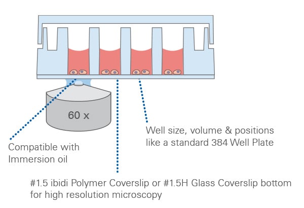
The µ-Slide 32 Well integrates 32 separate wells on a thin, high-performance #1.5 ibidi Polymer Coverslip or #1.5H Glass Coverslip bottom. The planar, transparent coverslip ensures excellent imaging performance and—together with the selected surface—supports defined, reproducible cell adhesion. The ibiTreat (tissue culture-treated) surface has been designed for optimal cell adhesion on the #1.5 ibidi Polymer Coverslip bottom. This modification provides ideal growth conditions for nearly all types of adherent cells, eliminating the need for additional coatings.
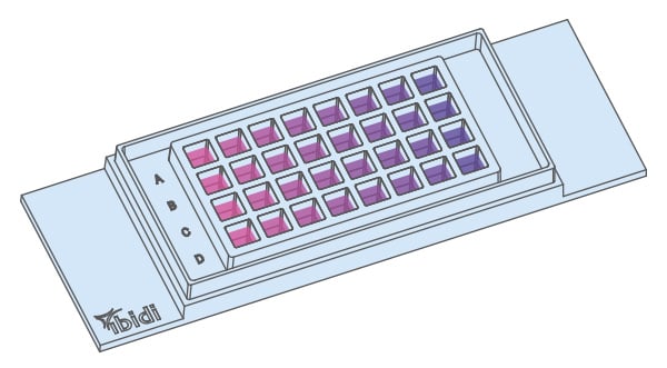
Multiple conditions can be run in parallel in the µ-Slide 32 Well.
The µ-Slide 32 Well at a Glance
| Product type | 32 well chambered coverslip for parallel cell-based assays and high-resolution microscopy |
| Best suited for | Assay development and optimization, immunofluorescence, parallel cell culture and treatment, high-content imaging, and high-resolution microscopy |
| Well geometry | 32 independent wells arranged according to the geometry of a standard 384 well format |
| Bottom and surface options | #1.5 ibidi Polymer Coverslip with ibiTreat or Untreated surface; #1.5H Glass Coverslip Bottom |
| Volume and growth area per well | 50 µl; 0.12 cm² |
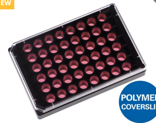
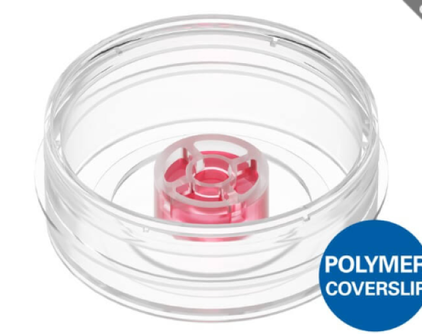
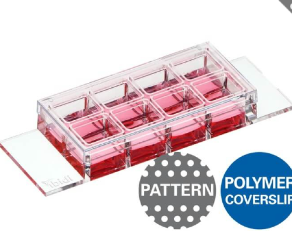
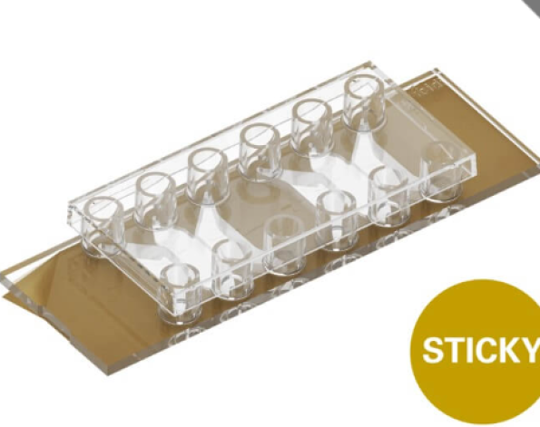
The ibidi Chambered Coverslip Variety
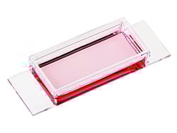 | 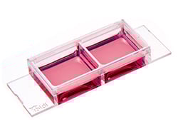 | 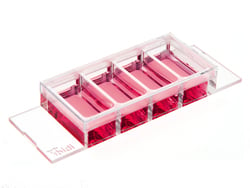 | 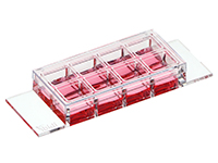 | 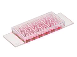 | 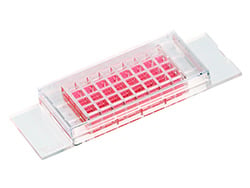 | |
| 1 Well | 2 Well | 4 Well | 8 Well high | 18 Well | 32 Well | |
| Volume per well | 3 ml | 1.5 ml | 700 µl | 300 µl | 100 µl | 50 µl |
| Growth area per well | 10.6 cm² | 5.1 cm² | 2.5 cm² | 1.0 cm² | 0.34 cm² | 0.12 cm² |
The ibidi Chambered Coverslips for cell culture, immunofluorescence, and high-end microscopy provide high flexibility in well size, well volume, and bottom materials. They are available in several formats, ranging from 1 large well to 32 small wells per µ-Slide. All microscopy slides are simple to use for immunofluorescence or live cell imaging assays without any coverslip handling. Available with either a #1.5 ibidi Polymer Coverslip or #1.5H Glass Coverslip bottom.
For a detailed comparison of coverslip materials, optical properties, and compatibility, visit the ibidi Material Comparison.
Frequently Asked Questions About the µ-Slide 32 Well
Why Is the Well Size of the µ-Slide 32 Well the Same as in a 384 Well Plate?
The well size, well-to-well distance, and working volume of the µ-Slide 32 Well are designed to match the dimensions of a high-throughput 384 well plate, such as the ibidi µ-Plate 384 Well or µ-Plate 384 Well Clear. This enables easy transfer of protocols and results from the µ-Slide 32 Well to a 384 Well screening platform without major protocol adaptations.
In brief, it combines the well geometry of a 384 Well imaging plate with the handling convenience of a slide.
When Should I Choose the µ-Slide 32 Well or a 384 Well Plate?
Choose the µ-Slide 32 Well when you want to compare multiple experimental conditions while minimizing cell and reagent consumption. The well geometry of the µ-Slide 32 Well matches a 384 well geometry facilitating the transfer of established protocols to 384 well plates (e.g., the ibidi µ-Plate 384 Well or µ-Plate 384 Well Clear). Choose a 384 well plate for automated liquid handling, high-content imaging, and high-throughput screening.
| Feature | µ-Slide 32 Well | 384 well plate |
| Well size | 3.5 mm | 3.4–3.6 mm (typically) |
| Working volume | 50 µl | 50 µl (typically) |
| Coverslip bottom | #1.5 ibidi Polymer Coverslip (different surfaces available) #1.5H Glass Coverslip (uncoated and untreated) | Polystyrene, glass coverslip, or polymer coverslip |
| Number of wells | 32 | 384 |
| Handling | Manual pipetting | Manual or automated liquid handling |
| Format | Microscopy slide | ANSI/SLAS microplate |
| Readout | High-resolution research microscope | High-content imaging platform |
Can I Use a Multichannel Pipette With the µ-Slide 32 Well?
Yes. The standard well positions in the µ-Slide 32 Well allow the use of multichannel pipettes.
How Does the µ-Slide 32 Well Simplify Assay Development?
The standard microscopy slide format of the µ-Slide 32 Well enables convenient handling and compatibility with common microscope stages and slide holders. Its low cell and reagent requirements help reduce sample consumption and costs during assay optimization. Once established, the assay can be transferred directly to the µ-Plate 384 Well for automated high-throughput applications.
Does the µ-Slide 32 Well Reduce Reagent Consumption?
Yes. The 32-well format enables many conditions with low working volumes, helping to conserve antibodies, dyes, and media while maintaining high imaging quality.
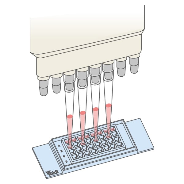
The wells of the µ-Slide 32 Well match the size and spacing of a standard 384 well plate, enabling easy transfer of cell models, protocols, and results from slide-based experiments to automated plate-based workflows.
Is the µ-Slide 32 Well Suitable for Live Cell Imaging?
Yes. The coverslip bottom delivers the optical quality required for long-term live cell imaging and time-lapse acquisition. For stable temperature, CO2, and humidity, combine the slide with a Stage Top Incubator.
Packaging Information
Pcs./Box: 15 (indiv. packed)
Coverslip: #1.5 Polymer: (180 µm +10/-5 µm), sterilized
Surface: ibiTreat: tissue culture-treated
Cat.No: 83206
Pcs./Box: 90 (indiv. packed)
Coverslip: #1.5 Polymer: (180 µm +10/-5 µm), sterilized
Surface: ibiTreat: tissue culture-treated
Cat.No: 83206-90
Pcs./Box: 120 (8 per tray)
Coverslip: #1.5 Polymer: (180 µm +10/-5 µm), sterilized
Surface: ibiTreat: tissue culture-treated
Cat.No: 83206-120
Pcs./Box: 15 (indiv. packed)
Coverslip: #1.5 Polymer: (180 µm +10/-5 µm), sterilized
Surface: Untreated
Cat.No: 83201
Pcs./Box: 90 (indiv. packed)
Coverslip: #1.5 Polymer: (180 µm +10/-5 µm), sterilized
Surface: Untreated
Cat.No: 83201-90
Pcs./Box: 120 (8 per tray, 15 trays)
Coverslip: #1.5 Polymer: (180 µm +10/-5 µm), sterilized
Surface: Untreated
Cat.No: 83201-120
Pcs./Box: 15 (indiv. packed)
Coverslip: #1.5H Glass: (170 µm +/-5 µm) D 263 M Schott glass, sterilized
Surface: Untreated
Cat.No: 83207
Pcs./Box: 90 (indiv. packed)
Coverslip: #1.5H Glass: (170 µm +/-5 µm) D 263 M Schott glass, sterilized
Surface: Untreated
Cat.No: 83207-90
Pcs./Box: 120 (8 per tray)
Coverslip: #1.5H Glass: (170 µm +/-5 µm) D 263 M Schott glass, sterilized
Surface: Untreated
Cat.No: 83207-120