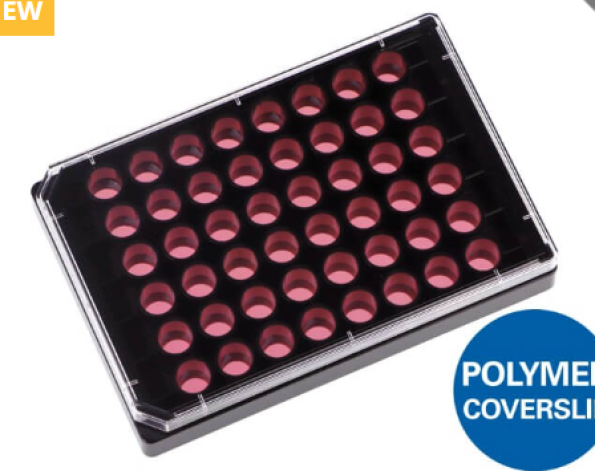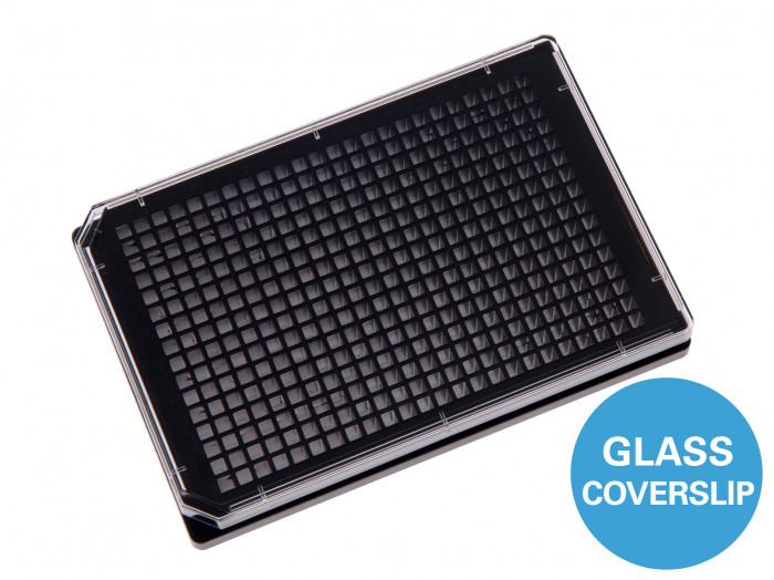
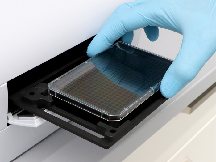
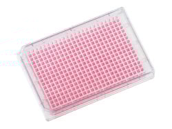
เพลท 384 หลุมสีดำสำหรับงานเพาะเลี้ยงเซลล์และการถ่ายภาพวิเคราะห์เซลล์ประสิทธิภาพสูง มาพร้อมตัวเลือกก้นหลุมแบบ Polymer Coverslip หรือ Glass Coverslip ที่ให้ความเรียบและคุณภาพเชิงแสงระดับสูง รองรับการถ่ายภาพด้วยกล้องจุลทรรศน์ความละเอียดสูงได้อย่างมีประสิทธิภาพ รูปแบบมาตรฐาน ANSI/SLAS ช่วยให้ใช้งานร่วมกับระบบอัตโนมัติและงาน High-Throughput Screening ได้อย่างราบรื่น ขณะที่ผนังหลุมสีดำช่วยลดการรบกวนของสัญญาณฟลูออเรสเซนซ์ระหว่างหลุม เพื่อเพิ่มความน่าเชื่อถือของผลการวิเคราะห์.
คุณสมบัติเด่น
- แผ่นเพลทสีดำชนิด 384 หลุม ออกแบบสำหรับการทดสอบแบบเซลล์ (Cell-based Assays) และงานถ่ายภาพวิเคราะห์เซลล์ความละเอียดสูง (High-Content Imaging)
- มีตัวเลือกก้นหลุมแบบ ibidi Polymer Coverslip #1.5 หรือ Glass Coverslip #1.5H ซึ่งให้ความเรียบทั้งภายในหลุมและทั่วทั้งแผ่นเพลทในระดับดีเยี่ยม เหมาะสำหรับการถ่ายภาพด้วยกล้องจุลทรรศน์ความละเอียดสูง
- ประกอบด้วย 384 หลุมทรงสี่เหลี่ยม ในมาตรฐาน ANSI/SLAS (SBS) Format รองรับระบบจ่ายสารอัตโนมัติ (Automated Liquid Handling) และงานคัดกรองตัวอย่างปริมาณสูง (High-Throughput Screening)
- ผนังหลุมสีดำช่วยลดการรบกวนของสัญญาณฟลูออเรสเซนต์ระหว่างหลุม (Well-to-Well Crosstalk) ทำให้ได้ผลการถ่ายภาพฟลูออเรสเซนซ์ที่แม่นยำยิ่งขึ้น
A black 384 well plate for cell-based assays and high-content imaging
- Choice of #1.5 ibidi Polymer Coverslip or #1.5H Glass Coverslip Bottom, with excellent inner well and whole plate flatness for high-resolution microscopy
- 384 square wells in ANSI/SLAS (SBS) format for automated liquid handling and high-throughput screening
- Black walls reduce well-to-well crosstalk for fluorescence microscopy
Miniaturized Cell-Based Assays With the µ-Plate 384 Well
The µ-Plate 384 Well is a black 384 well plate for cell culture, high-content imaging, and automated screening. Its square wells combine a flat, optically clear coverslip bottom with a growth area of 0.11 cm² and a recommended volume of 50 µl per well. Available bottom options include a #1.5 ibidi Polymer Coverslip with ibiTreat or Untreated surface, and a #1.5H Glass Coverslip Bottom. The ANSI/SLAS format supports parallel imaging-based assays in manual and automated workflows.
More Conditions per Plate, Smaller Volumes per Assay
The 384 well format enables parallel comparison of compound concentrations, treatments, controls, and biological replicates. The 16 × 24 layout provides four times as many wells as a 96 well plate, with a 4.5 mm well spacing for automated handling. A recommended volume of 50 µl per well supports assay miniaturization while maintaining direct optical access for quantitative microscopy.
Immunofluorescence and Protein Localization
The µ-Plate 384 Well supports high-throughput immunofluorescence directly in the plate. Cell cultivation, fixation, staining, washing, and imaging take place in the same wells, without transferring cells to separate coverslips. Fluorescence intensity, protein localization, and cell morphology provide image-based readouts for comparing antibody panels, staining conditions, and treatment responses.

Fluorescence microscopy of immunostained Rat1 fibroblasts in the µ-Plate 384 Well Glass Bottom. Cells were stained for F-actin (green), alpha-Tubulin (red), and nuclei (DAPI, blue) and imaged using a 60x objective.
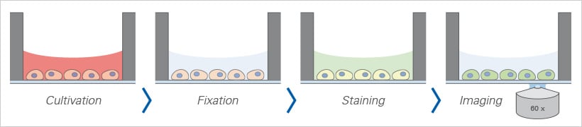
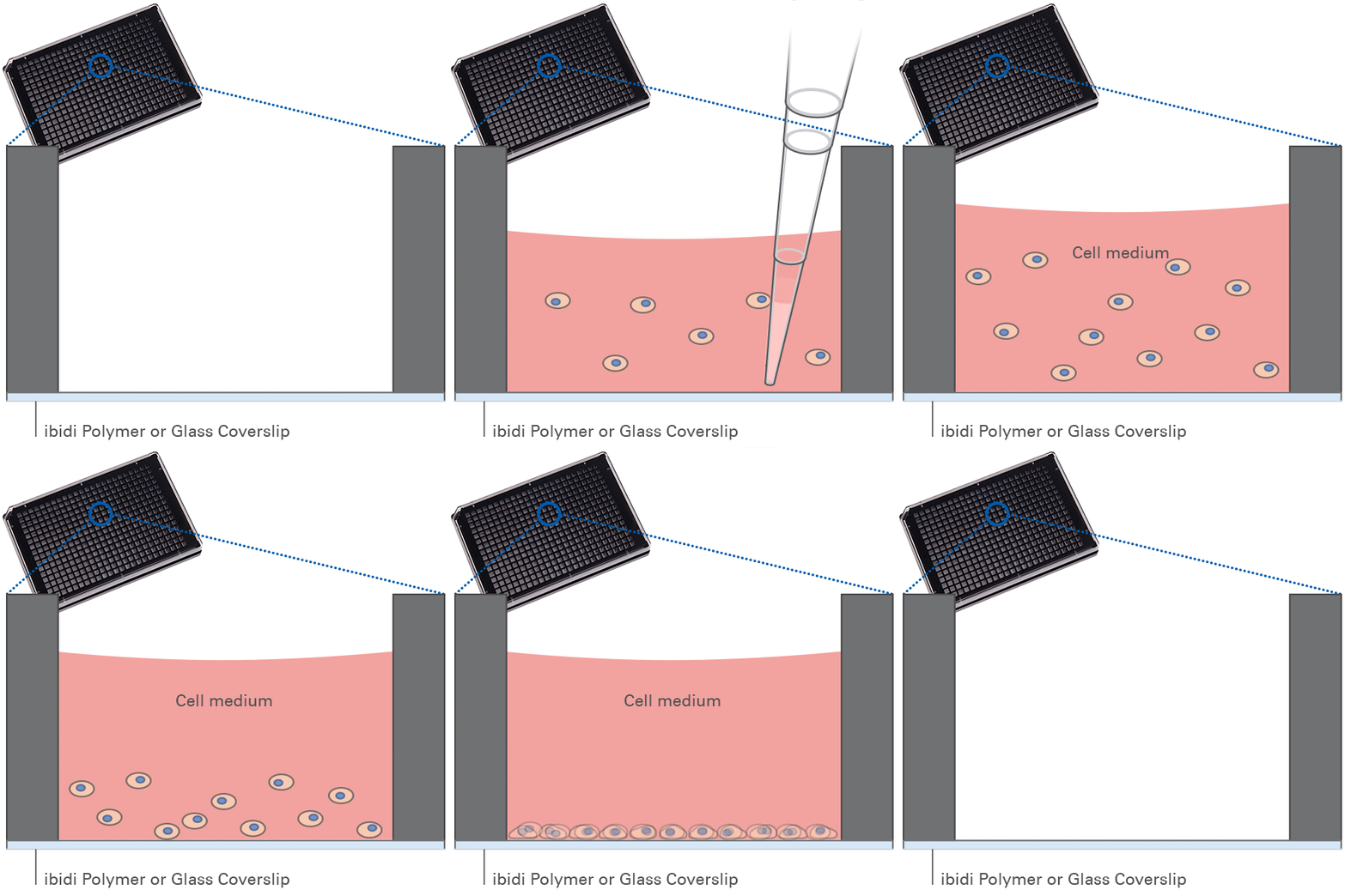
Example immunofluorescence workflow in the µ-Plate 384 Well: cell cultivation, fixation, staining, and imaging can be performed directly in the same plate.
Live Cell Imaging and Time-Lapse Studies
Live cell imaging enables monitoring of cell morphology, proliferation, reporter expression, and treatment responses. The coverslip bottom enables direct observation on inverted microscopes. Compatible environmental control, such as an ibidi Stage Top Incubator, supports long-term time-lapse studies. Humidity control and medium handling adapted to the small culture volumes help maintain suitable culture conditions.
Advanced Microscopy and Quantitative Readouts
The plate supports widefield fluorescence, confocal microscopy, and automated high-content screening (HCS). The #1.5H Glass Coverslip Bottom is recommended for demanding TIRF, super-resolution microscopy, and single-molecule workflows, subject to compatibility with the optical setup. Compatible fluorescence plate readers provide an additional option for well-level assay readouts.
Specifications
| Outer dimensions (l × w) | 127.7 × 85.5 mm |
| Number of wells | 384 |
| Height with/without lid | 17.2 / 15.0 mm |
| Well-to-well distance | 4.5 mm |
| Single well dimensions (l × w) | 3.4 × 3.4 mm |
| Single well depth | 12.7 mm (88401, 88406) 12.8 mm (88407) |
| Volume per well | 50 µl |
| Growth area per well | 0.11 cm² |
| Coating area per well using 50 µl | 0.8 cm² |
| Bottom material | #1.5 ibidi Polymer Coverslip (different surfaces available) #1.5H Glass Coverslip (uncoated and untreated) |
For technical drawings and details, please see:
Instructions µ-Plate 384 Well (PDF)
Instructions µ-Plate 384 Well Glass Bottom (PDF)
Technical Features
Precision, Quality, and Biocompatibility
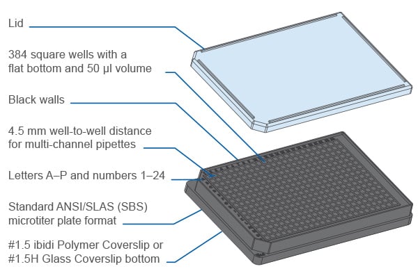
- µ-Plate (microtiter plate) in ANSI/SLAS (SBS) format
- Ready-to-use for lab automation and high-throughput with robotics, fluorescence scanners, and plate readers
- 384 square wells with standard numbering (letters A–P and numbers 1–24)
- Excellent inner well flatness and whole plate flatness
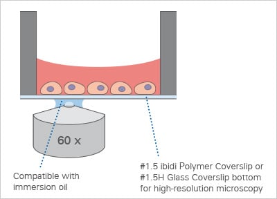
- #1.5 ibidi Polymer Coverslip or #1.5H Glass Coverslip bottom with excellent optical quality
- No autofluorescence due to black walls
- Compatible with staining and fixation solutions
- Compatible with immersion oil
- Made from non-cytotoxic, biocompatible materials
- Sterile with single packaging
Product Versions
- With #1.5 ibidi Polymer Coverslip (different surfaces available) or #1.5H Glass Coverslip Bottom
- Also available with transparent walls: µ-Plate 384 Well Clear
How Does the µ-Plate 384 Well Work?
The µ-Plate 384 Well combines 384 independent culture wells with a permanently integrated, thin coverslip bottom. Cells are cultured and imaged on the same surface, with optical access from below for inverted microscopy. The black walls separate neighboring wells and help reduce fluorescence crosstalk. The selected surface determines cell attachment: ibiTreat supports adherent cell culture, while Untreated Polymer and Glass Bottom versions allow application-specific coating workflows.
µ-Plate 384 Well at a Glance
| Product type | Black 384 well cell culture and imaging microplate |
| Best suited for | Miniaturized cell-based assays, high-content imaging, immunofluorescence, and automated screening |
| Well geometry | 384 square wells in a 16 × 24 array, each measuring 3.4 × 3.4 mm, with a flat, clear coverslip bottom |
| Bottom and surface options | #1.5 ibidi Polymer Coverslip with ibiTreat or Untreated surface; #1.5H Glass Coverslip Bottom, uncoated and untreated |
| Recommended volume and growth area per well | 50 µl; 0.11 cm² |
| Format | ANSI/SLAS (SBS) microplate format; 4.5 mm well spacing |
The ibidi µ-Plate Range
 |  | 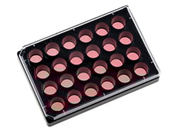 | 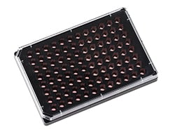 | 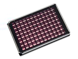 | 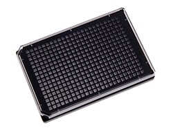 |  | |
| 6 Well | 12 Well | 24 Well | 96 Well Round | 96 Well Square | 384 Well | 384 Well Clear | |
| Bottom |   | | | | | | |
| Volume / well | 5 ml | 3 ml | 1 ml | 200 µl | 300 µl | 50 µl | 50 µl |
| Growth area / well | 9.1 cm² | 3.6 cm² | 1.54 cm² | 0.3 cm² | 0.56 cm² | 0.11 cm² | 0.11 cm² |
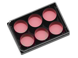
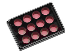
The ibidi µ-Plates are specifically designed for cell culture, immunofluorescence, and high-end microscopy. They offer flexibility in well size, well volume, and bottom materials. The range includes formats from 6 to 384 wells. All ibidi multiwell plates are user-friendly and ideal for multiplexing and high-throughput applications, providing high optical quality and compatibility with robotic systems. Most µ-Plates are available with either a #1.5 ibidi Polymer Coverslip or #1.5H Glass Coverslip bottom.
Packaging Information
| Cat. No. | Packaging Unit | Packaging Dimensions (l × w × h) | Shipping Weight |
|---|---|---|---|
| 88401, 88406, 88407 | 15 (indiv. packed) | 301 × 173 × 151 mm | 1.4 kg |
| 88401-90, 88406-90, 88407-90 | 90 (indiv. packed) | 505 × 319 × 244 mm | 8.7 kg |