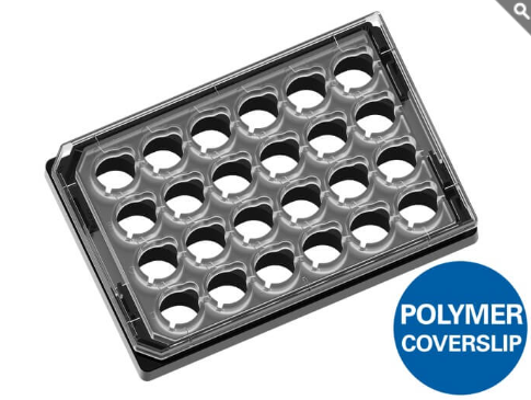
คุณสมบัติเด่น
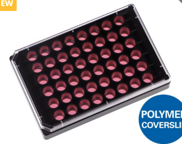
- เพลท 24 หลุม ออกแบบสำหรับการถ่ายภาพเซลล์ที่เพาะเลี้ยงบนเมมเบรนพรุน (Cell Culture Inserts with Porous Membranes) ได้อย่างมีประสิทธิภาพสูง
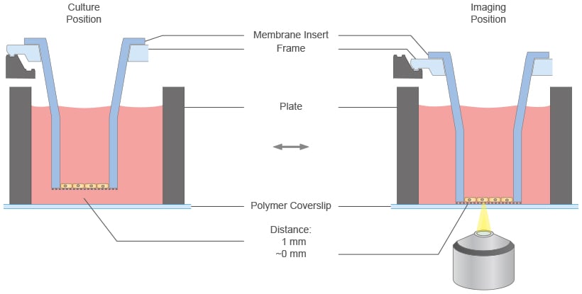
- มาพร้อมกลไก Two-Position Mechanism ที่ช่วยให้สามารถเข้าถึงและถ่ายภาพด้วยกล้องจุลทรรศน์ได้โดยไม่ต้องถอดเมมเบรนอินเสิร์ตออกจากเพลท*
- ก้นหลุมแบบ #1.5 Polymer Coverslip Bottom ที่มีความเรียบสูง ช่วยให้ได้ภาพคุณภาพเยี่ยมสำหรับการวิเคราะห์เซลล์บนเมมเบรนหรือวัสดุรองรับที่ยอมให้สารผ่านได้ (Permeable Supports)
- รองรับการใช้งานร่วมกับ Cell Culture Inserts จากหลายผู้ผลิต ได้แก่ Greiner, SARSTEDT, SABEU และ Millipore
Membrane Insert Assays, Designed for Imaging
The µ-Plate 24 Well for Membrane Inserts combines standard 24-well membrane inserts with direct access for high-quality microscopy. The two-position mechanism allows the inserts to be moved between cell culture and imaging positions, enabling direct observation through the optical bottom without removing the inserts or interrupting the experiment. It is ideal for barrier and transport studies, migration and invasion assays, co-culture models, and air–liquid interface (ALI) cultures.
Air–Liquid Interface Models
Culture epithelial cells under physiologically relevant air–liquid interface conditions to study differentiation, barrier formation, and tissue-specific functions.
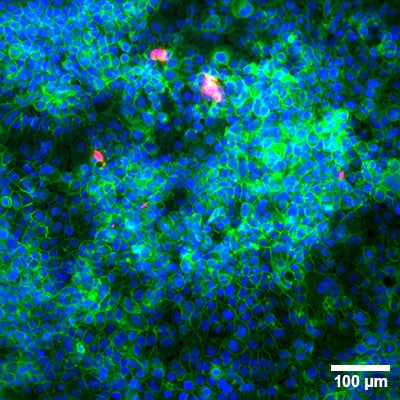
Bronchial epithelial cells (16HBE14o) cultured for 12 days at the air-liquid interface (ALI) on a membrane insert (Greiner; 0.4 µm pores) inside the µ-Plate 24 Well for Membrane Inserts. Nuclei are stained with DAPI (blue), F-actin with phalloidin-iFluor 488 (green), and mucus with Anti-Mucin 5AC conjugated to Atto 594 (red). Widefield fluorescence 20x.
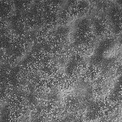
Phase contrast image (4x) of bronchial epithelial cells (16HBE14o) cultured for 12 days at the air-liquid interface (ALI) on Greiner inserts with 0.4 µm pores inside the µ-Plate 24 Well for Membrane Inserts.
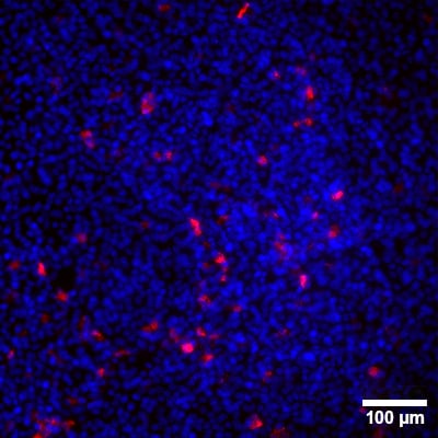
Primary human airway epithelial cells (hAEC) cultured for 20 days at the air-liquid interface (ALI) on the apical side of a membrane insert. Cell nuclei: DAPI (blue) and mucus produced by goblet cells: anti-mucin anibody (Atto 594, red). Widefield fluorescence microscopy 20x.
Co-Culture and Apical–Basal Cell Organization
Cultivate different cell types on opposite sides of a permeable membrane to investigate cell–cell communication, paracrine signaling, and multicellular interactions.
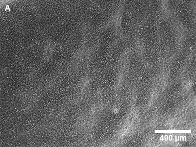
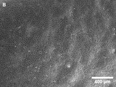
Co-culture system of hAECs on the apical side (A) and HUVECs on the basal side (B) of a Greiner insert with 0.4 µm pore size. Phase contrast, 4x.
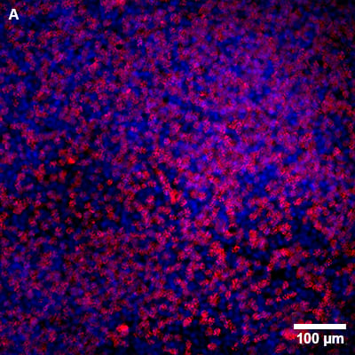
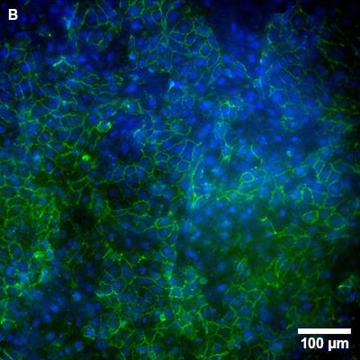
Co-culture of primary human airway epithelial cells (hAECs), grown on the apical side (A) for 20 days, and human umbilical vein endothelial cells (HUVECs), seeded on the basal side (B) on day 13. Cell nuclei: DAPI (blue), HUVECs: anti-CD31 (Alexa Fluor 488, green), and cilia of hAECs: anti-acetylated tubulin (Atto 594, red). Widefield fluorescence 20x.
High-Resolution Imaging
Capture detailed fluorescence images of cells cultured on membrane inserts, from widefield screening to high-resolution imaging with high-magnification objectives.
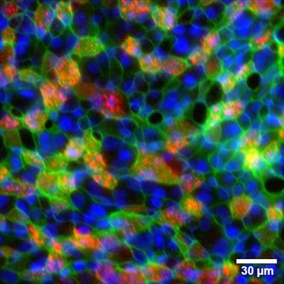
hAECs cultured for 20 days at the air-liquid interface (ALI) on the apical side of a membrane insert. Cell nuclei: DAPI (blue), F-actin: phalloidin-iFluor 488 (green), and cilia: anti-acetylated tubulin antibody (Atto 594, red). Widefield fluorescence microscopy with a 60x oil immersion objective.
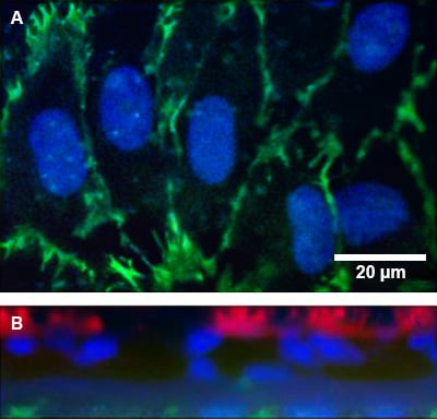
Fluorescence microscopy of a co-culture of primary human airway epithelial cells (hAECs), grown on the apical side for 20 days, and human umbilical vein endothelial cells (HUVECs), seeded on the basal side on day 13 in a Greiner insert (0.4 µm pores) within a µ-Plate 24 Well for Membrane Inserts. Cell nuclei were stained with DAPI (blue), HUVECs with anti-CD31 (Alexa Fluor 488, green), and the cilia of hAECs with anti-acetylated tubulin (Atto 594, red). Images show a single plane of the basal side (HUVECs) (A) and a z-stack cross-section of hAECs (apical) and HUVECs (basal) (B), acquired using a Nikon microscope with a 60x oil immersion objective.
Further Applications
The flexible membrane insert format supports a broad range of cell-based assays beyond the examples shown above.
Transmigration, Invasion, and Chemotaxis
Study immune cell transmigration, tumor cell invasion, and directional migration across porous membranes in response to chemical gradients.
Barrier, Permeability, and Transport Studies
Analyze epithelial and endothelial barrier function, molecular permeability, and transport across cellular layers under defined apical–basal conditions.
Drug Response and Automated Analysis
Combine membrane-based cell models with treatment studies and automated imaging for systematic comparison of multiple conditions in a standardized 24-well format.
Specifications
| Outer dimensions (l × w) | 127.7 × 85.5 mm |
| Number of wells | 24 |
| Height with/without lid | 20.7 / 18.6 mm |
| Compatible Membrane Inserts* | See list below |
| Well-to-well distance | 18.0 mm |
| Well clearance | 0.52 mm |
| Imaging distance Culture position Imaging position | 0 mm 1 mm |
| Single well depth | 9.3 mm |
| Volume per well | 1 ml |
| Growth area per well | 2.11 cm² |
| Coating area per well using 1 ml** | 4.7 cm² |
| Bottom material | #1.5 ibidi Polymer Coverslip (ibiTreat, tissue culture-treated) |
* Membrane inserts are not included with this product.
** Without consideration of a membrane insert.
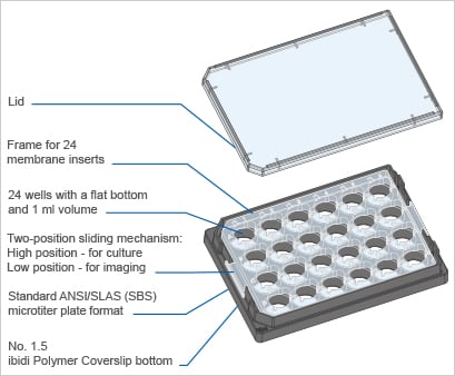
Technical drawings and details are available in the Instructions (PDF).
Technical Features
Automation
ANSI/SLAS (SBS) standard format suitable for automation and fluorescence scanners. Accommodates up to 24 cell culture inserts.*
Two-position Sliding Mechanism
Compatible with lab automation (robotic grippers).
- Culture position – Standard insert position (1 mm distance); recommended for cell culture.
- Imaging position – Moves the insert onto the coverslip (~0 mm distance); recommended for microscopy.
Compatibility
Compatible with 24-well-sized inserts from Greiner, SARSTEDT, SABEU, and Millipore; see compatibility list for details.
Staining and Microscopy
Excellent inner well flatness and whole plate flatness due to the #1.5 ibidi Polymer Coverslip. Compatible with staining and fixation solvents and immersion oils.
Material Properties and Handling
No autofluorescence, non-cytotoxic, and biocompatible in sterile single packaging.
* Membrane inserts are not included with this product.
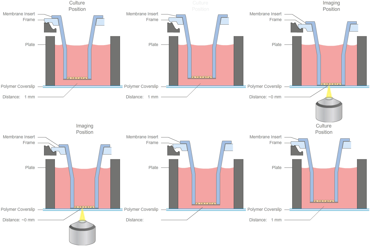
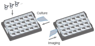
Use up to 24 inserts per plate and cultivate or image your cells by pushing the frame to the appropriate position. See list below for details on compatibility.
How Does the µ-Plate 24 Well for Membrane Inserts Work?
What Is the Principle of the µ-Plate 24 Well for Membrane Inserts?
The ibidi µ-Plate 24 Well for Membrane Inserts features a two-position sliding mechanism that enables easy switching between cultivation and high-resolution imaging. By this innovative design, the membrane can be put close to the ibidi Polymer Coverslip Bottom for optimal microscopy, and then can be manually returned to the standard hanging position for continued cell culture—ensuring seamless integration with commonly used inserts.
Which Membrane Inserts Are Compatible?
The plate is available in versions compatible with 24-well-sized inserts from Greiner Bio-One (ThinCert), SARSTEDT (TC-Inserts), SABEU (cellQART), or Millipore (Millicell). For details and catalog numbers, see the compatibility list on this page.
Which Applications Is the Plate Designed for?
Typical applications include transmigration and invasion assays, chemotaxis assays through porous membranes, barrier integrity and permeability studies (epithelial/endothelial), air-liquid interface (ALI) culture, apical–basal polarity studies, and microscopy-based readouts such as live cell imaging and immunofluorescence staining.
Can I Perform High-Resolution Imaging Without Removing the Inserts?
Yes. The imaging position reduces the distance between the membrane and the optical bottom, enabling improved access for high-resolution objectives. You can then return the plate to the culture position for continued cultivation using the same insert.
Compatibility List
The µ-Plate 24 Well for Membrane Inserts supports 24-well-sized membrane inserts from leading brands, including Millipore, Greiner Bio-One, SARSTEDT, and SABEU.
See the insert compatibility list for details:
| ibidi Product Name | Manufacturer | Compatible Catalog Numbers | |
| 82436 | µ-Plate 24 Well for Membrane Inserts Greiner/SARSTEDT | Greiner Bio-One | Greiner® ThinCert® Cell Culture Inserts: 662640, 662641, 662610, 662630, 662631, 662638 |
| SARSTEDT | SARSTEDT TC-Inserts: 83.3932.040, 83.3932.041, 83.3932.101, 83.3932.300, 83.3932.500, 83.3932.800 | ||
| 82446 | µ-Plate 24 Well for Membrane Inserts SABEU | SABEU | SABEU cellQART® Cell Culture Inserts: 932 04 02, 932 10 02, 932 30 02, 932 50 02, 932 80 02, 932 04 12, 932 10 12, 932 30 12, 932 50 12, 932 80 12, 932 04 22 |
| 82456 | µ-Plate 24 Well for Membrane Inserts Millipore | Millipore | Millicell Hanging Cell Culture Inserts: PTHT24H48, PCHT24H48, PLRP24H48, PTRP24H48, PTSP24H48, PCSP24H48, PTMP24H48, PTEP24H48, PCEP24H48 |
Please note that only the above-mentioned products are compatible and support the full functionality of the µ-Plate 24 Well for Membrane Inserts. Other insert products are not compatible. Membrane inserts are not included with this product.
Advantages Compared to a Standard 24 Well Plate
Porous membrane-based cell culture inserts have become standard tools in cell and tissue culture due to their versatility and ease of use. Membrane inserts, also known as cell culture inserts, hanging inserts, permeable supports, or porous membranes, are well-established and widely utilized in many cell culture assays and experimental setups. Typical applications include co-culture, transmigration and invasion assays, air-liquid interface (ALI) cultures, chemotaxis, drug transport and barrier function studies, tissue engineering, and 3D culture models such as spheroids and organoids. However, standard multiwell plates often limit microscopy performance due to:
- Thick plastic bottoms and a significant distance between the membrane and plate bottom, that restrict the use of high-resolution objectives.
- High autofluorescence, interfering with fluorescence-based imaging.
To overcome these limitations, the ibidi µ-Plate 24 Well for Membrane Inserts bridges the gap by enabling seamless transitions between cultivation and imaging. The unique design allows researchers to position the membrane with cells close to the high-quality ibidi coverslip for live cell or endpoint microscopy, then return it to the hanging position for continued cultivation. This two-position sliding technology provides optimal optical conditions for microscopy while maintaining full compatibility with commonly used membrane inserts.
ibidi µ-Plate 24 Well for Membrane Inserts (Imaging Position) | Standard 24 well plate with membrane inserts | |
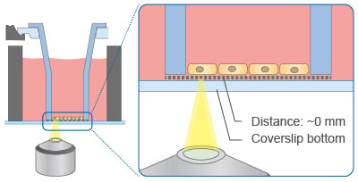 | 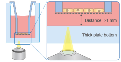 | |
| Bottom material | Coverslip bottom: high resolution, no autofluorescence | Thick plate bottom: low resolution, high autofluorescence |
| Imaging | Minimal distance: maximum microscopy access | Large distance: no microscopy access |
- Superior Imaging Performance – Designed for high-resolution and fluorescence microscopy with cell culture inserts.
- Uncompromised Experimental Conditions – Maintain your existing protocols, SOPs, and workflows without the need for adaptation.
- Compatible with Standard Inserts – Supports 24-well-sized membrane inserts from leading brands, including Millipore, Greiner Bio-One, SARSTEDT, and SABEU. See the insert compatibility list for details.
| Order No. | Packaging Unit | Packaging Dimensions (w × l × h) | Shipping Weight |
| 82436, 82446, 82456 | 15 (indiv. packed) | 301 × 173 × 151 mm | 1.3 kg |