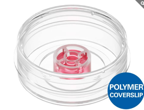
Silicone Insert สำหรับสร้าง Gel Matrix และ Cellular Interface
- ออกแบบสำหรับสร้าง Cellular Gel Interface พร้อมการเข้าถึงและแลกเปลี่ยนสารได้ทั้งด้าน Basal และ Apical ของเซลล์
- ดีไซน์พิเศษ ปราศจากเมมเบรน (Membrane-Free Design) ช่วยให้สามารถสังเกตและถ่ายภาพเซลล์ด้วยกล้องจุลทรรศน์ได้อย่างเต็มประสิทธิภาพ โดยไม่มีสิ่งกีดขวางในแนวการมองภาพ
- พร้อมใช้งานได้ทันทีร่วมกับ µ‑Dish 35 mm, high (ibiTreat) ช่วยลดขั้นตอนการเตรียมอุปกรณ์และเพิ่มความสะดวกในการทดลอง
จุดเด่น
- เหมาะสำหรับงานสร้างแบบจำลองเนื้อเยื่อ (Tissue Models) และการเพาะเลี้ยงเซลล์ในเจล 3 มิติ
- รองรับการศึกษาเซลล์ในสภาพแวดล้อมที่ใกล้เคียงสรีรวิทยาจริง
- สามารถเข้าถึงตัวอย่างได้ทั้งด้านบนและด้านล่างของชั้นเซลล์
- ให้คุณภาพการถ่ายภาพที่ยอดเยี่ยม เนื่องจากไม่มีเมมเบรนมารบกวนการสังเกตด้วยกล้องจุลทรรศน์
- ใช้งานง่ายและพร้อมเริ่มการทดลองได้ทันทีใน µ‑Dish 35 mm, high (ibiTreat)
A silicone insert designed for gel matrices and cellular interfaces
- Cellular gel interface with fluidic access to both basal and apical sides
- Unique, membrane-free design ensures optimal microscopy access to the cells
- Ready-to-use in the µ-Dish 35 mm, high, ibiTreat
Applications
The micro-Insert 3D is a biocompatible silicone insert used for creating cellular interfaces on or in gel matrices for:
- 3D cell culture with hydrogels (e.g., collagen or Matrigel)
- Live cell imaging of cells inside or on top of a hydrogel matrix
- Immunofluorescence staining
- Co-culture
- Transport assays
- Apical-basal cell polarity assays
- Cell barrier model assays with apical-basal gradients
- Cell migration assays (e.g., immune cell transmigration, tumor invasion, or metastasis)
Specifications
| Insert diameter | 12 mm |
| Insert height | 6 mm |
| Inner well diameter | 3 mm |
| Inner well volume | 25 µl |
| Outer well volume | 250 µl |
| Gel matrix volume* | 20 µl |
| Gel matrix height* | 0.8 mm |
| Gel matrix diameter* | 3 mm |
| Growth area on gel* | 0.07 cm2 |
| Material | Silicone |
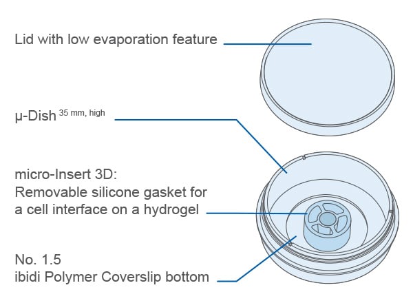
| Bottom material | ibidi Polymer Coverslip (µ-Dish 35 mm, high ibiTreat) |
*The gel matrix is not included with this product.
Technical Features
- 3 mm inner well geometry creates a flat gel surface when using 20 µl of gel*
- No artificial membranes — excellent optical properties for microscopy
- Compatible with various gel types, including Matrigel®, collagen, and others*
- Flexible assays with cells in 2D and 3D gel matrices
- Suitable for live cell imaging, fixation, and immunofluorescence staining
- Sterile with single packaging
- Ready-to-use in a µ-Dish 35 mm, high
*The gel matrix is not included with this product.
For technical drawings and details, please see:
Instructions micro-Insert 3D (PDF)
Product Benefits
The micro-Insert 3D can be used for applications in which porous membrane inserts are typically utilized. Instead of a porous membrane, the micro-Insert 3D uses a hydrogel to separate the well into an outer (Solution A) and an inner (Solution B) compartment. This membrane-free design allows improved microscopy access in phase contrast and fluorescence microscopy — ideal for long-term, high-resolution live cell imaging. Compared to porous membranes, the micro-Insert 3D enables more in vivo-like modeling for cell cultures: no hard surfaces, no artificial pores, and no rigid cellular interface. The unique design permits direct fluidic access to both the apical and basal sides of the cells and gel matrix.
The micro-Insert 3D has an adhesive underside that prevents leaking on smooth, dry surfaces, like microscopy slides and dishes. It can be ordered ready-to-use in our µ-Dish 35 mm, high (80496) or in a transport petri dish for self-insertion (80499). The micro-Insert 3D can easily be transferred to any labware using sterile tweezers.
- Save reagents due to low gel volume
- Mimics native in vivo conditions combined with excellent microscopy access (no artificial membranes)
- Physiologically relevant 3D assay approach with high flexibility

How Does the micro-Insert 3D Work?
What Is the Principle of the micro-Insert 3D?
The micro-Insert 3D is a bottomless silicone insert with an adhesive underside that is used to cast a small hydrogel (typically 20 µl) in the central inner well. This hydrogel separates the system into an outer and an inner compartment, enabling a membrane-free cell–gel interface for imaging and transport-style assays.
What Is the micro-Insert 3D Used for?
It is used for 3D cell culture and microscopy workflows where cells are cultured on or in a hydrogel matrix, including live cell imaging, immunofluorescence staining, barrier/polarity setups, migration/transmigration assays, and co-culture configurations with separate apical/basal compartments.
Is the Gel Matrix Included?
No. The gel matrix is not included. The micro-Insert 3D is compatible with commonly used hydrogels such as Matrigel® and Collagen Type I.
What Are the Advantages of the Cell-Gel Interface Compared to Classical Porous Membranes?
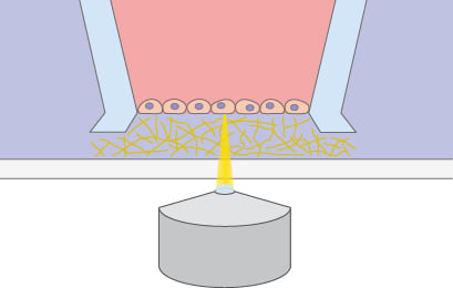 | 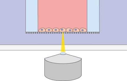 | |
| Cell-Gel Interface With the micro-Insert 3D | Cell Interface With a Membrane Insert | |
| Principle | Cells on hydrogel matrix | Cells on porous membrane |
| Preparation | Casting hydrogel of choice (e.g., ECM modeling) | Coating with protein of choice |
| Diffusion | Open for diffusion; tunable by choice of gel matrix | Limited diffusion; dependent on pore size, membrane thickness, and porosity |
| Mechanical properties | Soft gel matrix; physiological Stiffness and pore sizes defined by gel | Stiff polymer membrane; artificial Pore sizes defined by membrane |
| Chemical properties | Hydrogel can be remodeled; cell interaction possible | Membrane is inert; no biological interaction |
| Cell retrieval | Gel digestion, mechanical retrieval | Mechanical scraping, chemical trypsinization |
| Microscopy access | Good: direct microscopy access | Poor: membrane cutting necessary |
| Imaging quality | Hydrogels are well accessible by microscopy | Membranes restrict microscopy access |
Technical Possibilities
The micro-Insert 3D offers a range of assay possibilities by enabling flexible cell culture configurations. It is primarily used to create a cell layer on top of a hydrogel, forming a cell interface that divides the chamber into basal and apical compartments. Various modifications can also be implemented, such as embedding cells or spheroids/organoids within the gel matrix. Additionally, cells can be cultured in the outer well to support co-cultivation workflows.
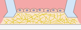
Cells on the gel
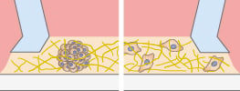
Cells in the gel
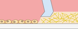
Cells outside the micro-Insert
Application Examples
Immunofluorescence
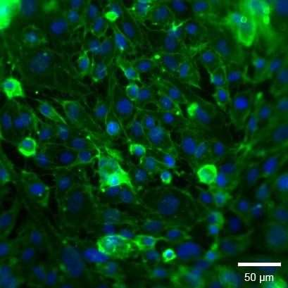
Widefield fluorescence microscopy of HUVECs on Collagen Type I, Rat Tail in the micro-Insert 3D. Cells were fixed after 48 h. The F-actin cytoskeleton was stained with Alexa Fluor 488-phalloidin (green) and nuclei with DAPI (blue). Image acquired on a Nikon microscope using a 20x objective lens.
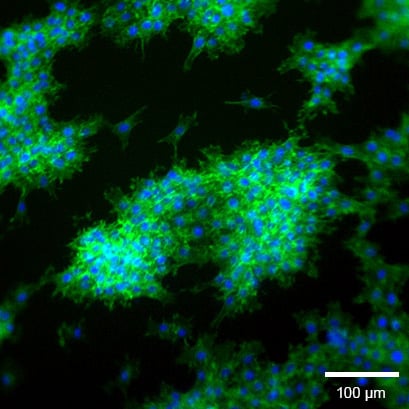
Widefield fluorescence microscopy of rat fibroblasts (Rat1) on Collagen Type I, Rat Tail in the micro-Insert 3D. Cells were fixed after 24 h. The F-actin cytoskeleton was stained with Alexa Fluor 488-phalloidin (green) and nuclei with DAPI (blue). Image acquired on a Nikon microscope using a 10x objective lens.
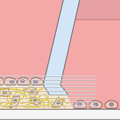
Volume 3D stack of human fibrosarcoma cells (HT-1080) in Collagen Type I, Rat Tail (3D cell culture) in the micro-Insert 3D. Cells were fixed after 72 h. The F-actin cytoskeleton was stained with Alexa Fluor 488-phalloidin (green) and nuclei with DAPI (blue). Image acquired on a Nikon microscope using a 20x objective lens.
Live Cell Imaging
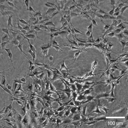
Phase contrast image of mouse fibroblasts (NIH3T3) on Collagen Type I, Rat Tail in the micro-Insert 3D. Image acquired after 24 h on an EVOS microscope using a 4x objective lens.
Potential Applications
The following examples are untested and are only meant to show potential product uses. ibidi still needs to verify these applications in-house, so we cannot provide specific protocols at this time.
Barrier
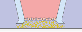
A cell monolayer is cultivated on a hydrogel matrix, creating a cellular barrier.
Migration
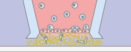
Transmigration of, for example, leukocytes through a cell monolayer.
Tumor Angiogenesis
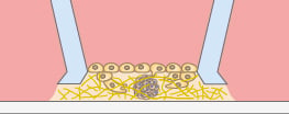
Sprouting of, for example, endothelial cells towards tumor cells.
Air-Liquid Interphase
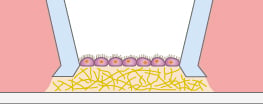
A monolayer of, for example, epithelial cells cultivated on a hydrogel matrix.
Co-Culture
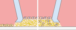
A cell monolayer is cultivated on a hydrogel matrix while a second cell type is present either inside the 3D matrix or as a 2D monolayer.
Advanced Co-Culture
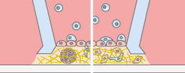
Transmigration of, for example, immune cells across a cell monolayer into a 3D gel matrix with embedded tumor cells.
Packaging Information
| Order No. | Packaging Unit | Packaging Dimensions (w × l × h) | Shipping Weight |
| 80496 | 30 (indiv. packed) | 305 × 140 × 92 mm | 0.3 kg |
| 80496-200 | 200 (indiv. packed) | 305 × 305 × 305 mm | 2.2 kg |