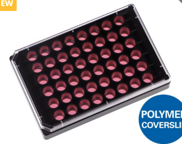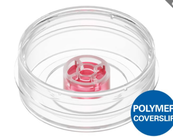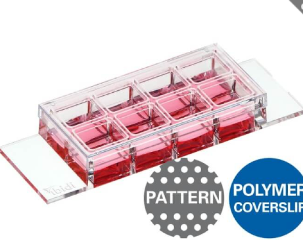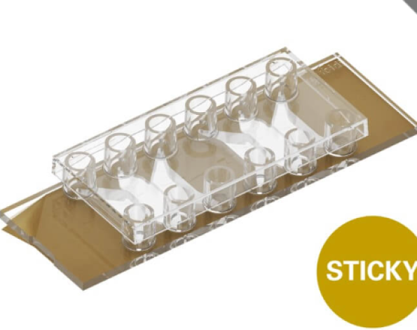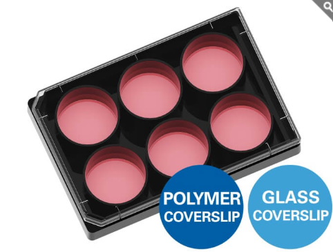
ibidi : Black 6-Well Plate ก้นใสและเรียบ สำหรับการเพาะเลี้ยงเซลล์ การย้อม IF และงานกล้องจุลทรรศน์
- แผ่นเพาะเลี้ยงเซลล์แบบ Multiwell ขนาด 6 หลุม มาพร้อมก้นหลุมที่เป็น #1.5 Polymer Coverslip หรือ #1.5H Glass Coverslip ให้คุณภาพทางแสงยอดเยี่ยม เหมาะสำหรับการถ่ายภาพด้วยกล้องจุลทรรศน์ความละเอียดสูง
- ออกแบบให้มีความเรียบสูงทั้งภายในแต่ละหลุมและทั่วทั้งแผ่นเพลต ช่วยเพิ่มความแม่นยำและความสม่ำเสมอในการทำ Automated Screening
- ผนังหลุมสีดำ (Black Walls) ช่วยลดการรบกวนของสัญญาณฟลูออเรสเซนซ์ระหว่างหลุม (Well-to-Well Crosstalk) ทำให้ได้ผลการวิเคราะห์ที่คมชัดและเชื่อถือได้มากขึ้น
จุดเด่น
- คุณภาพการมองภาพยอดเยี่ยม รองรับงาน Fluorescence Microscopy และ High-Resolution Imaging
- ก้นหลุมเรียบเป็นพิเศษ เหมาะสำหรับระบบคัดกรองอัตโนมัติ (Automated Screening)
- ผนังสีดำช่วยลดสัญญาณรบกวนระหว่างหลุม เพิ่มความแม่นยำของการวัดฟลูออเรสเซนซ์
- เหมาะสำหรับการเพาะเลี้ยงเซลล์ การย้อม Immunofluorescence (IF) และงานวิเคราะห์ด้วยกล้องจุลทรรศน์ขั้นสูง
Black 6 well plate with a flat and clear bottom for cell culture, IF, and microscopy
- Multiwell plate with #1.5 Polymer Coverslip or #1.5H Glass Coverslip bottom with excellent optics
- Cell culture plate with excellent inner well flatness and whole plate flatness for automated screening
- Microplate with black walls for low well-to-well crosstalk during fluorescence microscopy
Six Conditions in Parallel With High Cell Numbers
The µ-Plate 6 Well supports experiments requiring higher cell numbers, extended culture periods, or sufficient material for downstream analyses. The plate is optimized for cell culture and high-resolution imaging without transferring samples. Ideal for cell morphology studies, proliferation and migration assays, live cell imaging, and endpoint analyses such as immunofluorescence.
Immunofluorescence and Protein Localization
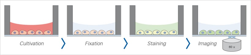
The ibidi µ-Plate 6 Well allows standard immunofluorescence protocols to be performed directly in the plate format. All steps: cell cultivation, fixation, staining, and imaging occur in the open well geometry. After staining, samples are imaged through the coverslip-like bottom using high-resolution microscopy.
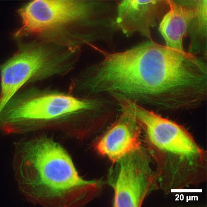
Widefield fluorescence microscopy of immunostained human umbilical vein endothelial cells (HUVEC) in the µ-Plate 6 Well ibiTreat. The F-actin cytoskeleton was stained with phalloidin (red), the cell nuclei were stained with DAPI (blue) and the alpha-Tubulin was stained with an anti-Tubulin antibody (green). The image was taken on a Nikon microscope using a 60x objective lens.
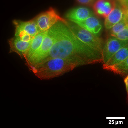
Widefield fluorescence microscopy of immunostained MCF-7 cells (human breast cancer) in the µ-Plate 6 Well Glass Bottom. The F-actin cytoskeleton was stained with phalloidin (red), the cell nuclei were stained with DAPI (blue) and the alpha-Tubulin was stained with an anti-Tubulin antibody (green). The image was taken on a Nikon microscope using a 60x objective lens.
Cell Cultivation and Imaging
- Cultivation and high-resolution microscopy of cells
- Parallel cell culture experiments with low cell numbers and reagent volumes
- High-sensitivity immunofluorescence stainings and protein localization studies
- Live cell imaging and time-lapse studies over extended time periods
- Large-scale gene and protein expression analysis, transfection assays
- Drug discovery studies, cytotoxicity assays, and toxicology screenings with clear visibility of cell morphology
Microscopy Techniques
- Brightfield, phase contrast, and widefield fluorescence microscopy for cell monitoring on inverted microscopes
- Automated, multiparametric high-content screening (HCS) and high-throughput screening (HTS) with plate readers
- Super-resolution microscopy (STED, (F)PALM, (d)STORM, SIM) and TIRF (best output with #1.5H Glass Coverslip)
- Confocal microscopy, two-photon microscopy, FRAP, FRET, FLIM, and LSFM
Specifications
| Outer dimensions (l × w) | 127.8 × 85.5 mm |
| Number of wells | 6 |
| Height with/without lid | 22.4 / 20.0 mm |
| Well-to-well distance | 37.5 mm |
| Single well diameter | 34.0 mm |
| Single well depth | 18.5 mm |
| Volume per well | 5 ml |
| Growth area per well | 9.1 cm² |
| Coating area per well using 5 ml | 14.9 cm² |
| Bottom material | #1.5 ibidi Polymer Coverslip (different surfaces available) #1.5H Glass Coverslip (uncoated and untreated) |
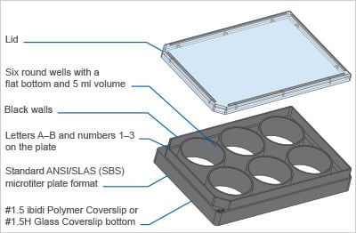
For technical drawings and details, please see:
Instructions µ-Plate 6 Well (PDF)
Instructions µ-Plate 6 Well Glass Bottom (PDF)
Technical Features
Precision, Quality, and Biocompatibility
- µ-Plate (microtiter plate) in ANSI/SLAS (SBS) format
- Ready-to-use for lab automation and high-throughput with robotics, fluorescence scanners, and plate readers
- Six round wells with standard numbering (letters A–B and numbers 1–3)
- Excellent inner well flatness and whole plate flatness
- #1.5 ibidi Polymer Coverslip or #1.5H Glass Coverslip bottom with excellent optical quality
- No autofluorescence due to black walls
- Compatible with staining and fixation solutions
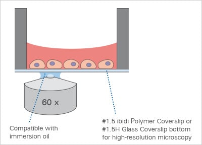
- Compatible with immersion oil
- Made from non-cytotoxic, biocompatible materials
- Sterile with single packaging
Product Versions
- With #1.5 ibidi Polymer Coverslip (different surfaces available) or #1.5H Glass Coverslip Bottom
How Does the µ-Plate 6 Well Work?
What Is the Principle of the µ-Plate 6 Well?
The µ-Plate 6 Well provides six independent wells on a thin, high-performance #1.5 ibidi Polymer Coverslip or #1.5H Glass Coverslip bottom. The flat, transparent imaging bottom delivers excellent optical quality and—together with the chosen surface—supports defined, reproducible cell adhesion. The ibiTreat (tissue culture-treated) surface has been designed for optimal cell adhesion on the #1.5 ibidi Polymer Coverslip bottom. This modification provides ideal growth conditions for nearly all types of adherent cells, eliminating the need for additional coatings.
What Is the µ-Plate 6 Well Used for?
With its standard microplate footprint, the µ-Plate 6 Well is suited for parallel cell culture and imaging workflows, such as immunofluorescence staining, brightfield and fluorescence microscopy, TIRF, and live cell imaging on inverted microscopes. Typical use cases include condition screens, dose–response experiments, and side-by-side controls in one plate.
Is the µ-Plate 6 Well Compatible With Automated Handling?
Yes. The microplate format fits common stage inserts and plate holders and is well suited for automated liquid handling, enabling reproducible, medium-throughput assays. It is ready-to-use for lab automatization and high-throughput with robotics, fluorescence scanners, and plate readers.
Is the µ-Plate 6 Well Suitable for Live Cell Imaging?
Yes. The coverslip bottom provides the optical performance required for long-term live cell imaging. For stable temperature, CO2, and humidity, use it together with a Stage Top Incubator.
How Do I Conduct Immunofluorescence Stainings With the µ-Plate 6 Well?
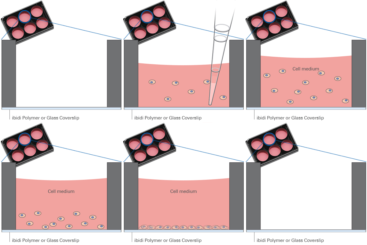
The µ-Plate 6 Well is well suited for immunofluorescence (IF) assays. Six separate wells enable multiple conditions and replicates in one plate, with convenient washing and staining directly in the wells. The integrated #1.5 or #1.5H imaging bottom provides excellent optics for widefield fluorescence and confocal microscopy—no additional coverslip mounting or sample transfer is required.
The ibidi µ-Plates Variety
 | 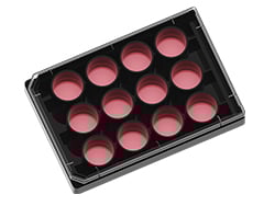 | 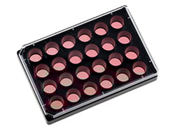 | 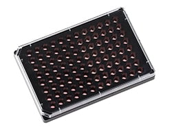 | 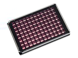 | 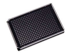 | 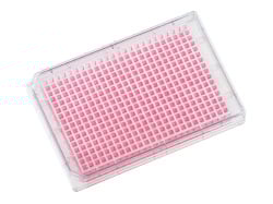 | |
| 6 Well | 12 Well | 24 Well | 96 Well Round | 96 Well Square | 384 Well | 384 Well Clear | |
| Bottom |   | | | | | | |
| Volume / well | 5 ml | 3 ml | 1 ml | 200 µl | 300 µl | 50 µl | 50 µl |
| Growth area / well | 9.1 cm² | 3.6 cm² | 1.54 cm² | 0.3 cm² | 0.56 cm² | 0.11 cm² | 0.11 cm² |
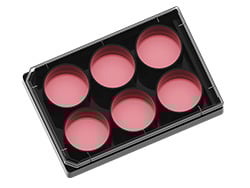
The ibidi µ-Plates are specifically designed for cell culture, immunofluorescence, and high-end microscopy. They offer flexibility in well size, well volume, and bottom materials. Available in various formats, these plates range from 6 to 384 wells per µ-Plate. All ibidi multiwell plates are user-friendly and ideal for multiplexing and high-throughput applications, ensuring top optical quality and compatibility with robotic systems.
Most µ-Plates are available with either a #1.5 ibidi Polymer Coverslip or #1.5H Glass Coverslip bottom—both offering excellent optical clarity for standard microscopy techniques such as brightfield, phase contrast, and confocal microscopy. The tissue culture-treated ibiTreat Polymer Coverslip supports the growth of most cell types without any need for additional coatings and eases the use in cell microscopy.
While differences between the ibidi Polymer and Glass Coverslips are minimal in standard microscopy workflows, the choice of material can significantly affect advanced techniques such as TIRF and super-resolution microscopy.
For detailed optical specs, refer to our Surface Material Guide or evaluate the different coverslip materials through our Free Sample Program.
| Order No. | Packaging Unit | Packaging Dimensions (w × l × h) | Shipping Weight |
| 80631, 80636, 80637 | 15 (indiv. packed) | 301 × 173 × 151 mm | 1.2 kg |
| 80631-90, 80636-90, 80637-90 | 90 (indiv. packed) | 505 × 319 × 244 mm | 7.0 kg |