กว่า 25 ปีแห่งนวัตกรรม ประสิทธิภาพ และความน่าเชื่อถือสำหรับห้องปฏิบัติการวิจัย
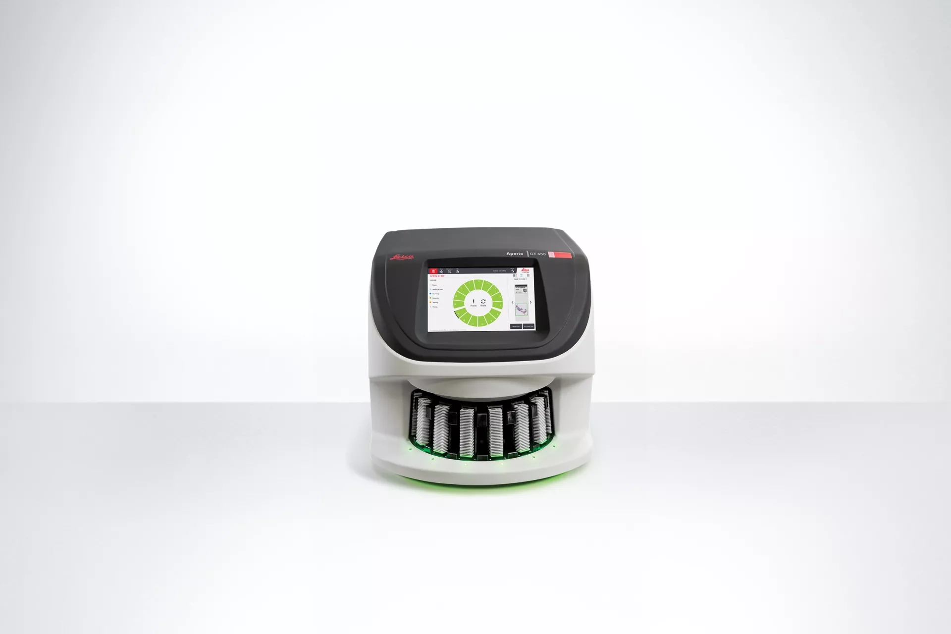
เครื่องสแกนสไลด์แบบดิจิตอล Aperio GT 450 ผสานเทคโนโลยีล้ำสมัยและนวัตกรรมที่ได้รับการพัฒนาอย่างต่อเนื่อง เพื่อมอบความยืดหยุ่นในการสแกนและระบบเวิร์กโฟลว์อัตโนมัติ ช่วยให้ห้องปฏิบัติการได้ผลลัพธ์ที่มีคุณภาพสูง รวดเร็ว และมีประสิทธิภาพ
คุณสมบัติและประโยชน์เด่นของ Aperio GT 450
- สิทธิบัตรมากกว่า 40 รายการ (40+ Patents)
รองรับความต้องการในการสแกนที่หลากหลาย พร้อมสนับสนุนเวิร์กโฟลว์อัตโนมัติเพื่อเพิ่มประสิทธิภาพการทำงาน - ความเร็วในการสแกนเพียง 32 วินาทีต่อสไลด์
สามารถสแกนได้สูงสุด 81 สไลด์ต่อชั่วโมงที่กำลังขยาย 40x* ช่วยเพิ่มปริมาณงานและลดระยะเวลาการทำงาน - Manual Scan
สามารถปรับตั้งค่าการสแกนและสแกนซ้ำได้ตามต้องการ เพื่อให้ได้คุณภาพภาพที่ดีที่สุดสำหรับแต่ละตัวอย่าง - Extended Focus
ผสานข้อมูลจากหลายระนาบโฟกัสเข้าด้วยกันเพื่อสร้างภาพที่คมชัดยิ่งขึ้น พร้อมช่วยลดความต้องการพื้นที่จัดเก็บข้อมูล - Default Calibration Point
ช่วยรักษาคุณภาพของภาพให้สม่ำเสมอ ลดปัญหาการเกิดแถบเส้น (Image Striping) ในภาพสแกน - รองรับมาตรฐาน DICOM
รองรับการสแกนที่กำลังขยาย 20x และ 40x พร้อมฟังก์ชัน Z-Stack เพื่อสร้างภาพที่ละเอียด คมชัด และเหมาะสำหรับงานวิเคราะห์เชิงลึก - Aperio iQC Software
ใช้เทคโนโลยีปัญญาประดิษฐ์ (AI) ในการตรวจจับสิ่งรบกวนหรือข้อบกพร่องของภาพ (Artifacts) ช่วยให้การตรวจสอบคุณภาพและการทบทวนภาพเป็นไปอย่างรวดเร็วและมีประสิทธิภาพยิ่งขึ้น
oven Technology for Researchers
Effortless scanning, exceptional results with excellent image quality, innovative technology and seamless integration – all driven towards faster turnaround time and accuracy for your important projects

Manual Scan
Delivering researchers with an efficient solution for variability in slide preparation, Manual Scan provides:
- Faster slide delivery with immediate rescanning from the same location.
- Quick resolution of image issues by lab technicians including faintly stained IHC slides, small tissue fragments, low density tissues etc.
- High-quality slides from challenging tissues.
- Streamlined workflow and improved lab efficiency.
Extended Focus
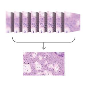
Experience an efficient imaging solution that improves image quality while reducing storage needs. Extended Focus features include:
- Viewing all cells in focus on a single image, eliminating the need to scroll through layers.
- Smaller file sizes than Z-Stack images speed up file transfers, improving collaborative activities.
- Ideal in applications where thick specimens are viewed, which make the extended depth of field of images an issue to address.
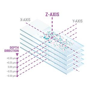
DICOM with 20x,40x and Z-Stack
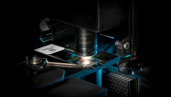
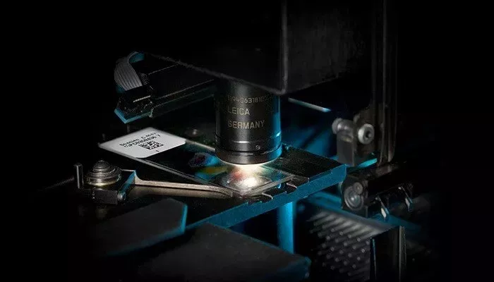
Aperio GT 450 uses a high-performance objective manufactured by Leica Microsystems, which has produced world-class optics since 1847. Leica optics deliver exceptional image quality
- Available alongside .svs, DICOM enables image clarity, improved collaboration and a streamlined workflow.
- 20x and 40x options for clear and detailed views of samples
- Z-Stacking enables the review of slide samples at varied heights
Proven Technology for Lab Managers
Deliver rapid results with confidence.
The Aperio GT 450 is simple to operate and helps you complete scanning quickly and easily, so you can focus on other tasks in the lab: 15 racks of 30 slides (450 slides total) can be loaded directly from HistoCore SPECTRA CV Coverslipper into the Aperio GT 450. The slides are then automatically scanned at 81 slides per hour at 40x for a 15mm x 15mm area.
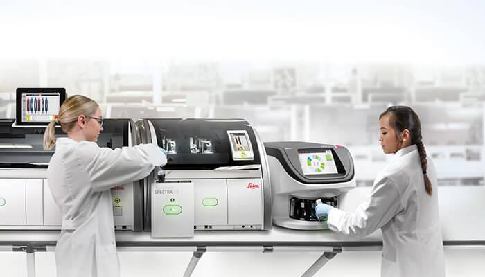
The products, shown above, HistoCore SPECTRA Workstation, in conjunction with Aperio GT 450 are for Research Use Only (RUO).
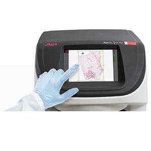
Manual Scan
Providing lab technicians with an intuitive and efficient scanning solution, the benefits of Manual Scan include:
- Easy-to-learn functionality that technicians can quickly navigate. Choose the scan area, set focus points and calibrate with ease.
- Enhanced control and confidence throughout the scanning process.
- Ability to successfully scan challenging slides with greater ease.
- Improved workflow efficiency.
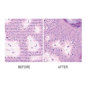
Default Calibration Point
The Aperio GT 450 Default Calibration Point reduces the chance of striping from the start, allowing lab managers to maintain image clarity and avoid corrective measures. This ensures consistently high-quality results with no extra effort.
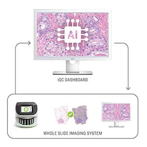
Aperio iQC software
Optional Aperio iQC software enhances image quality control, ensuring confidence before scans are delivered to the researcher.
- AI-enabled detection of whole slide image (WSI) artifacts, with notifications and a quality control dashboard to flag issues and recommend actions.
- Algorithms designed to analyze WSIs and identify potential artifacts.
- Automatic assessment to flag substandard slides, optimizing manual review and lab technicians’ time.
Proven Technology for IT Professionals
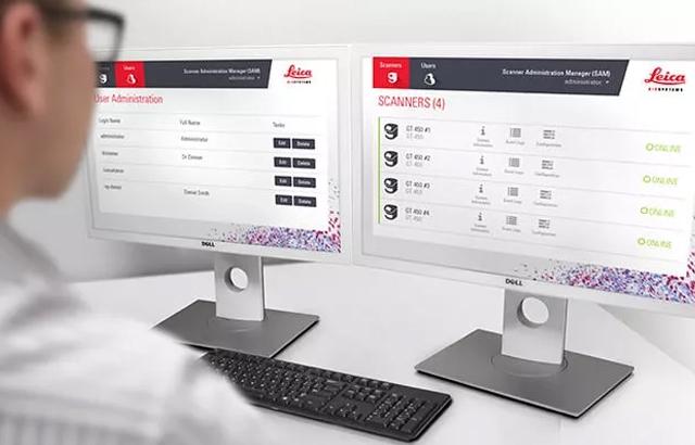
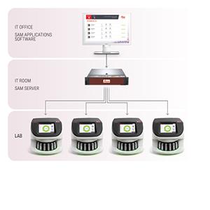
The Aperio GT 450 offers fast, secure, and flexible IT architecture. A centralized Scanner Admin Manager (SAM) server and dedicated software provides you with a data management solution that can remotely set up and monitor multiple Aperio GT 450s at a time.*
- Comprehensive cybersecurity including encryption and login controls
- Centralized scanner management to set up and monitor multiple scanners
- Two 10GB network connections on the SAM server
- Scalable set up for hub and spoke configurations
- HL7 compatible for easy integration into LIS , LIMS or PACS
- Supports dedicated logins for each Aperio GT 450 scanner
SAM Server
No more workstations needed. A centralized Scanner Admin Manager (SAM) enables IT professionals to scale up Digital Pathology operations securely and efficiently. SAM server and software allow IT managers to set up and monitor Aperio GT 450 scanners via the network, including dashboard status views, PIN controls and log out times.
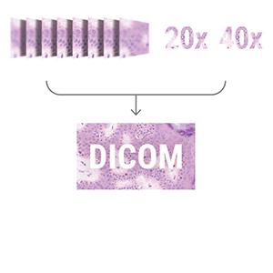
DICOM File
The DICOM format enhances compatibility for Z-Stacking, 20x and 40x magnification, offering high-quality images that can be easily shared and accessed across different systems. Now available alongside .svs, DICOM enables image clarity, improved collaboration and a streamlined workflow.
Image Quality Tested by Scientists for Scientists
(for best viewing results we recommend the Aperio Viewing Station which includes a calibrated monitor)
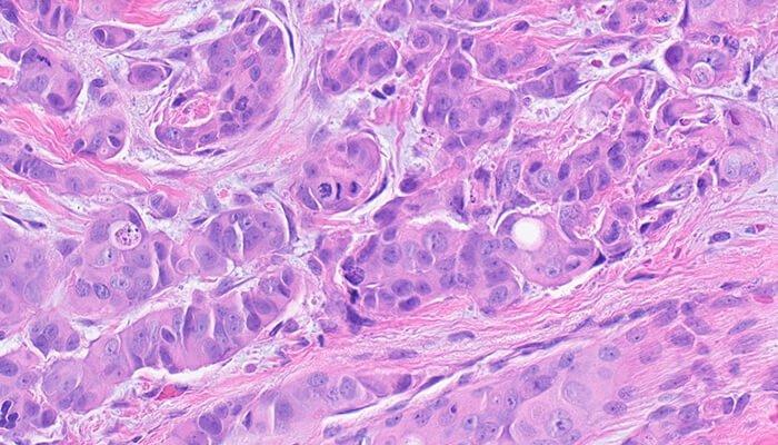
Mitotic Events (Breast)
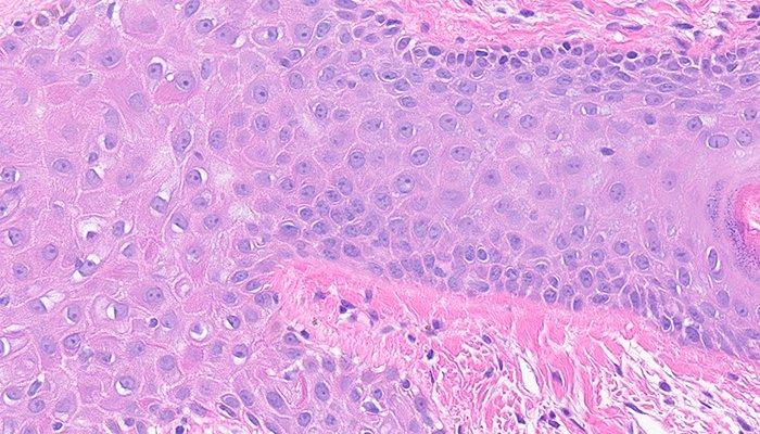
Intercellular Bridges
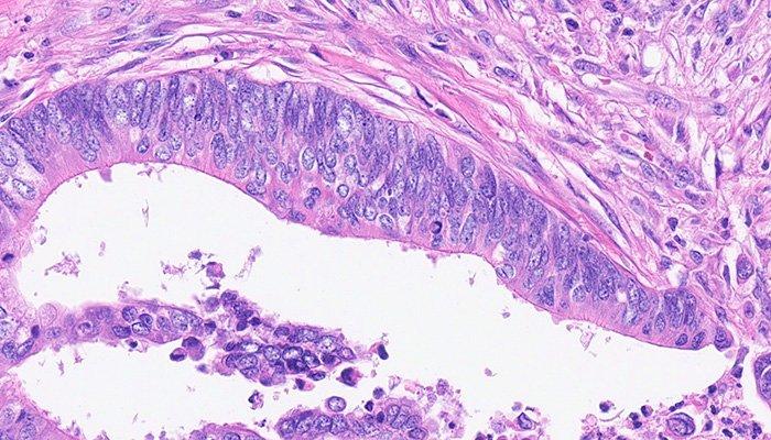
Chromatin
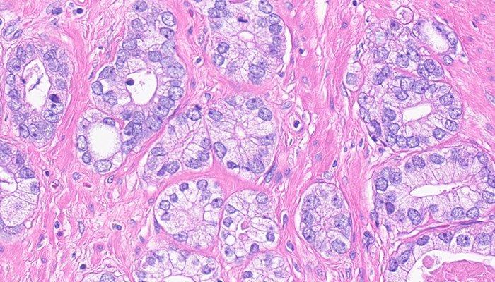
Nucleoli
Precision, Potential to Scale, and Peace of Mind
Digital pathology scan speeds have not always been fast enough at 40x magnification to keep up with high-volume scanning (120k+ slides per year). Real-Time Focusing (RTF)** offers a potential solution to this problem. It’s a novel method to capture a digital image that combines an imaging line sensor and a focusing line sensor.
- While scanning, the focusing line sensor receives focusing data of the tissue.
- Proprietary algorithms determine the best focus value.
- Then, each best focus value is treated like a control loop with the mechanics, feeding the best focus value to the objective height on the fly.
- The positioning adjustments of the objective are performed continuously (in real time) in order to enable the imaging line sensor to capture the image at its best possible focus. Hence, real time focusing.
**US Patent no. 9,841,590
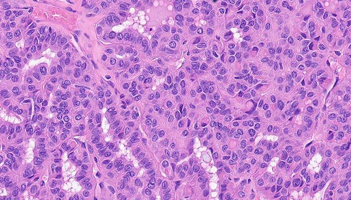
Cellular Groves (Thyroid)
Excellent Image Quality with Custom-Made Leica Objective
The high-performance objective included with the Aperio GT 450 is specifically designed to maximize field of view for high speed digital pathology scanning. Most objective are designed similar to the human eye: round, which focuses light in the center and limits field of view. The objective on the Aperio GT 450 includes an extra-wide flat field correction that enables a much larger field of view (1mm) that can accommodate extremely large digital images for fast scans and 0.26 um/pixel resolution at 40x magnification*.
*Available in 20x magnification.