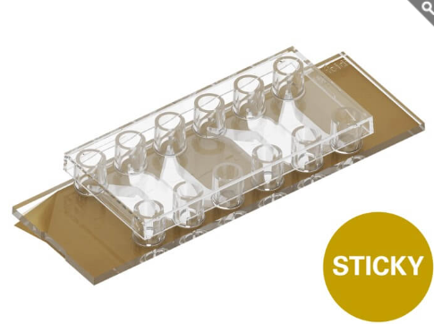
คุณสมบัติเด่น
- สไลด์ไมโครแชนแนลแบบกาว (Microchannel sticky-Slide) สำหรับงาน Organ-on-a-Chip, Tissue Models และ 3D Bioprinting
- แพลตฟอร์มไมโครฟลูอิดิกที่รองรับการพิมพ์ชีวภาพสามมิติ (Direct 3D Bioprinting) ภายในแชนแนลโดยตรง รวมถึงการสร้างโครงสร้างและลวดลาย 2D/3D ด้วยเทคนิคการกระตุ้นด้วยแสง (Light-Induced Microfabrication)
- แชนแนลแบบไม่มีก้นหลุม (Bottomless Channel Slide) พร้อมพอร์ตเชื่อมต่อมาตรฐาน Luer ช่วยให้เชื่อมต่อกับระบบปั๊มและควบคุมการไหลของของเหลวได้อย่างราบรื่น
- มีความยืดหยุ่นสูงในการใช้งาน โดยด้านกาวสามารถติดตั้งเข้ากับแผ่น Coverslip หรือวัสดุฐานรองชนิดต่าง ๆ ได้ตามความต้องการของงานวิจัย
Microchannel sticky-Slide for organ-on-a-chip assays, tissue models, and 3D bioprinting
- Microfluidic platform for direct 3D bioprinting or light-induced 2D/3D microfabrication into the channel
- Bottomless channel slide with fluidic access by standard Luer ports for seamless pump integration
- Highly versatile: sticky side can be mounted on a variety of coverslips or other bottom materials
Applications
Tissue and Organ-on-a-Chip Models
- Bioprinted functional architectures: Create complex 3D tissue architectures through precise bioprinting of cells and hydrogels directly into the central workspace.
- Tissue and organ models: Build micro-scale models of tissues (e.g., organ-on-a-chip) for studying cell behavior, drug effects, or tissue regeneration.
- Custom 3D structures: Fabricate microchannels, bifurcations, or flow barriers by bioprinting or—in combination with a coverslip bottom—by photoinduced polymerization using photocrosslinkable hydrogels.
Microfluidics
- High-precision microfluidics: When mounted onto a bottom material, seamlessly integrate the ibidi Pump System to control perfusion of microchannels and 3D structures.
- Modeling interstitial flow: Mimic the movement of fluids through tissues to study how cells sense and respond to shear stress. Bioprinted porous or fibrous structures allow researchers to tailor flow resistance and matrix permeability to match native tissue.
- Gradient formation: Generate gradients in 3D by dynamic perfusion.
- Dynamic microenvironments: Use controlled perfusion to maintain cell viability and mimic changing physiological conditions.
Microscopy Techniques
- Brightfield, phase contrast, and widefield fluorescence microscopy for cell monitoring on inverted microscopes
- Live cell imaging and time-lapse studies over extended time periods
- Confocal microscopy, two-photon microscopy, FRAP, FRET, FLIM, and LSFM
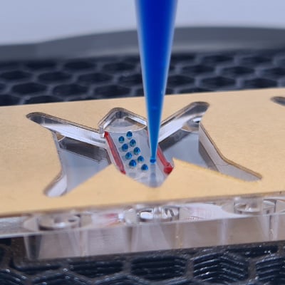
The sticky-Slide Tissue Engineering on a 3D printer. The open access enables flexible sample preparation (e.g., application of bioinks and cells by bioprinting).

Philipp’s Trick:
What’s an Easy Way to Seal and Perfuse 3D Bioprinted Structures?
Find out on our Tips and Tricks page!
Specifications
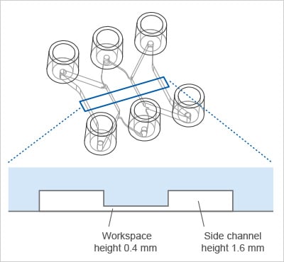
| Outer dimensions (w × l) | 25.5 × 75.5 mm |
| Adapters/reservoirs | Female Luer |
| Volume workspace | 36 µl |
| Height workspace | 0.485 mm |
| Growth area workspace | 0.77 cm2 |
| Coating area workspace | 1.56 cm2 |
| Volume side channels | 140 µl each |
| Height side channels | 1.685 mm |
| Growth area side channels | 0.64 cm2 each |
| Coating area side channels | 2.00 cm2 each |
| Volume per reservoir | 60 µl |
| Material | Polymer |
| Bottom | None (bottomless) |
For technical drawings and details, please see:
Instructions sticky-Slide Tissue Engineering (PDF)
Technical Features
Precision, Quality, and Biocompatibility
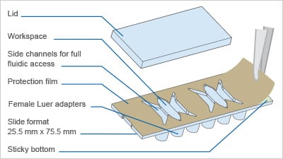
- Slide with 2 workspaces, each flanked by parallel channels
- Sticky bottom for easy mounting onto ibidi coverslips, standard glass microscope slides, or other materials
- Open workspace for direct 3D bioprinting (before mounting)
- Compatible with bioinks, fluidic photocrosslinkable hydrogels, or extracellular matrices
- Compatible with staining and fixation solutions
- Compatible with immersion oil
- Made from non-cytotoxic, biocompatible materials
- Sterile with single packaging
- Fully compatible with high-resolution fluorescence microscopy when combined with the #1.5 ibidi Polymer or #1.5H Glass Coverslip
- Ideal in combination with the ibidi Micro Illumination System
- Standard Luer ports: compatible with multiple flow and perfusion setups (e.g., ibidi Pump System)
Product Versions
- Also available: µ-Slide Tissue Engineering, a closed microchannel slide with #1.5 ibidi Polymer Coverslip bottom
How Does the sticky-Slide Tissue Engineering Work?
What Is the Principle of the sticky-Slide Tissue Engineering?
The sticky-Slide Tissue Engineering provides a flexible platform for designing and studying complex 2D and 3D tissue models under controlled fluidic conditions. The slide consists of a central workspace flanked by parallel channels. Before mounting the sticky-Slide onto a bottom material, the workspace is openly accessible for direct bioprinting. After mounting, photocrosslinkable hydrogels can be polymerized into defined patterns using the ibidi Micro Illumination System or similar systems. The design of the slide allows for seamless integration with the ibidi Pump System for controlled perfusion, simulating physiological flow and nutrient transport.
What Is the sticky-Slide Tissue Engineering Used For?
The sticky-Slide Tissue Engineering is designed to be combined with bioprinters to create advanced 2D and 3D cell culture experiments that require a realistic tissue environment and fluid control, such as tissue and organ modeling, flow and perfusion studies, and gradient formation.
How Can I Bioprint Into the sticky-Slide Tissue Engineering?
The sticky-Slide Tissue Engineering can be easily placed onto the build platform of a 3D bioprinter. Its fully open workspace provides direct access for printing bioinks, hydrogels, and cells. This enables flexible sample preparation—such as extrusion of structured scaffolds, printing of perfusable channels, or the placement of multi-material tissue constructs—before the slide is sealed onto a coverslip or another bottom material.
Is the sticky-Slide Tissue Engineering Compatible With the ibidi Pump System?
Yes. After mounting the sticky-Slide Tissue Engineering on a bottom material, it can be easily connected to pump systems such as the ibidi Pump System via the Luer adapters for precise control of perfusion rates and flow profiles. This setup allows you to simulate physiological conditions such as blood flow, interstitial flow, or dynamic shear stress. The channels can be perfused in parallel to achieve complex flow scenarios.
Is the sticky-Slide Tissue Engineering Suitable for Microscopy and Live Cell Imaging?
The sticky-Slide Tissue Engineering should be mounted on an optical high-quality coverslip, such as the #1.5 ibidi Polymer or #1.5H Glass Coverslip for microscopy. Physiological conditions can be maintained with the ibidi Stage Top Incubators or other incubators for live cell imaging.
Do I Need the ibidi Micro Illumination System to Use the sticky-Slide Tissue Engineering?
The sticky-Slide Tissue Engineering is designed for bioprinting of 3D structures directly into the workspace before mounting. However, after mounting onto a coverslip bottom, you can perform 2D or 3D photostructuring with the ibidi Micro Illumination System or other photoinduced technologies.
Application Example
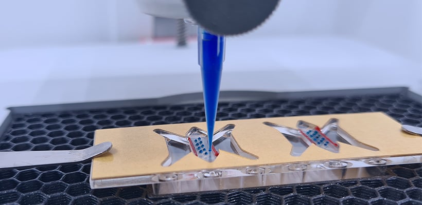
The sticky-Slide Tissue Engineering on a 3D printer. The open access enables flexible sample preparation (e.g., application of bioinks and cells by bioprinting).
Packaging Information
| Order No. | Packaging Unit | Packaging Dimensions (w × l × h) | Shipping Weight |
| 80238 | 15 (indiv. packed) | 150 × 130 × 110 mm | 0.2 kg |
| 80238-90 | 90 (indiv. packed) | 295 × 225 × 175 mm | 1.4 kg |
| 80238-120 | 120 (8 per tray) | 295 × 225 × 133 mm | 1.9 kg |