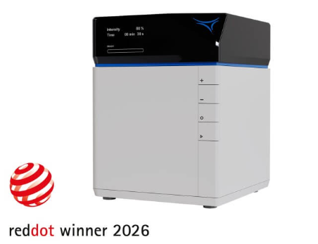
สร้างโมเดลเซลล์ขั้นสูงได้โดยตรงภายในห้องปฏิบัติการของคุณ
ด้วย ibidi Micro Illumination System ผู้ใช้งานสามารถควบคุมตำแหน่งและช่วงเวลาที่เซลล์ยึดเกาะ เคลื่อนที่ และจัดเรียงตัวเป็นเนื้อเยื่อได้อย่างแม่นยำ ช่วยให้สามารถสร้างแบบจำลองทางสรีรวิทยาระดับจุลภาคในหลอดทดลอง (microphysiological in vitro models) ที่มีความใกล้เคียงกับสภาพจริงทางชีววิทยามากขึ้น
เครื่องมือนี้เป็นแพลตฟอร์มแบบตั้งโต๊ะ (benchtop platform) ที่มีขนาดกะทัดรัด ใช้งานง่าย และนำเทคโนโลยีการสร้างโครงสร้างระดับจุลภาคด้วยแสง (light-induced microfabrication) เข้าสู่การใช้งานประจำวันในห้องปฏิบัติการ โดยไม่จำเป็นต้องใช้ห้องคลีนรูม (cleanroom)
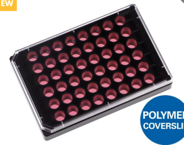
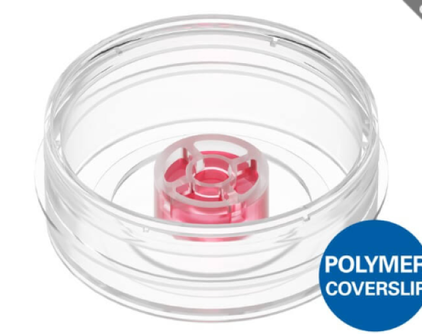
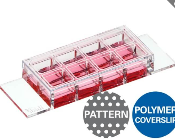
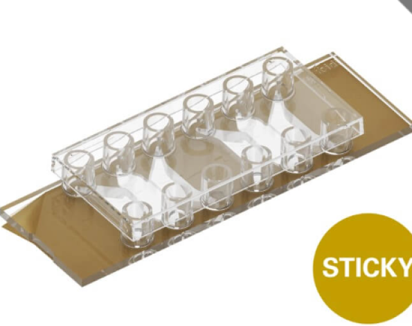
ระบบรองรับการใช้งานร่วมกับภาชนะเพาะเลี้ยงเซลล์แบบมีแผ่น coverslip เช่น µ‑Slides และ µ‑Dishes และรองรับเทคนิคทั้งแบบ 2 มิติ และ 3 มิติ (2D/3D) ที่หลากหลาย ได้แก่
- การสร้างลวดลายเซลล์ด้วยแสง (Photopatterning)
- การขึ้นรูปไฮโดรเจลระดับจุลภาค (Hydrogel Microstructuring)
- ปฏิกิริยา Photo-Click Chemistry
จึงเป็นโซลูชันที่ช่วยให้นักวิจัยสามารถพัฒนาแบบจำลองเซลล์และเนื้อเยื่อที่ซับซ้อน มีความแม่นยำ และมีความเกี่ยวข้องกับสภาวะทางชีววิทยาจริงได้อย่างสะดวกภายในห้องปฏิบัติการของตนเอง.
Build Advanced Cell Models Directly in Your Lab
With the ibidi Micro Illumination System, users can precisely control where and when cells attach, migrate, and organize into tissues, enabling the generation of relevant microphysiological in vitro models.
The instrument is a compact, user-friendly benchtop platform that brings light-induced microfabrication into everyday laboratories without the need for a cleanroom. Compatible with coverslip chambers like µ‑Slides and µ‑Dishes, it supports a broad range of 2D and 3D methods, including photopatterning, hydrogel microstructuring, and photo-click chemistry.
reate Perfusable Microfluidic Models
Hydrogel-based microchannels, bifurcations, and flow restrictions can be fabricated directly in ibidi Channel Slides. These structures support controlled perfusion, mechanical stimulation, and chemical gradient formation in lab-on-a-chip and organ-on-a-chip models.
Microfluidic Atherosclerosis Models
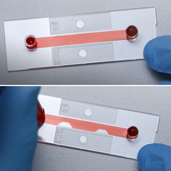
Flow obstacles in the µ-Slide I 0.4 Luer created with the ibidi Illumination System in a GelMA hydrogel. Top: normal channel of the µ-Slide I Luer filled with cell culture medium. Bottom: channel containing microstructures. Trapezoidal obstacles were created by crosslinking a GelMA hydrogel using the ibidi Micro Illumination System. HUVEC cells were then seeded to mimic a blood vessel with flow constrictions. Refer to Application Note 74 for the complete protocol.
Perfusable Hydrogel Tissue Models
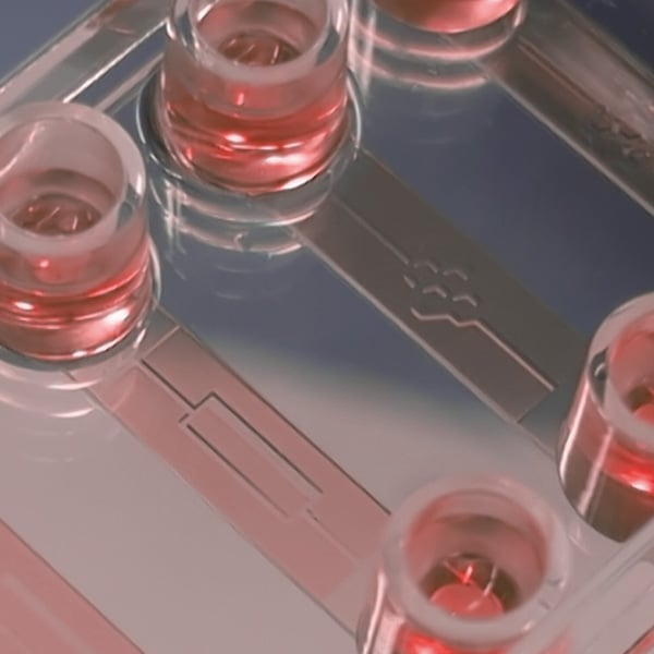
Microfabrication of a PEG-NB hydrogel, cross-linked with a cleavable peptide sequence directly in the µ-Slide VI 0.4 using the ibidi Micro Illumination System. Image shown in false color (original gel color: yellow; displayed as pink). Refer to Self-organized homogenization of flow networks for more information. Image courtesy of Charlott Leu, Soft Matter Physics, LMU, Munich.
Study Tissue Repair by Photo-Induced Wounding
Photo-induced wounding creates precisely defined cell-free areas in confluent monolayers. These reproducible wounds can be used to investigate cell migration, wound closure, endothelial repair, and tissue regeneration.
Fibroblast-Driven Monolayer Repair
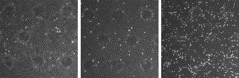
Optical wounding of rat fibroblast cells. Defined 200 µm areas were illuminated using the ibidi Micro Illumination System, resulting in the removal of living cells. Within 24 hours, the wounded areas were re-colonized by migrating cells, resulting in the formation of a new confluent monolayer. Refer to Application Note 76 for the complete protocol.
Perfused Endothelial Cell Migration and Wound Healing
Cell migration of endothelial cells under shear stress (10 dyn/cm²) in the µ-Slide VI 0.4. HUVECs were cultured under shear stress for 3 days before a 450 µm-wide band was optically wounded by UV irradiation using the ibidi Micro Illumination System. Perfusion was then resumed, and a time-lapse acquisition over 10 hours revealed that cells migrated from both sides to close the gap, thereby restoring a confluent monolayer. Refer to Application Note 76 for the complete protocol.
Control Cell Organization by Micropatterning
High-precision photopatterning enables spatially controlled surface functionalization and protein patterning to guide cell adhesion, migration, and organization. Photo click chemistry also allows defined spatial and temporal control of surface modifications.
Defined Cell Adhesion and Spatial Cell Organization
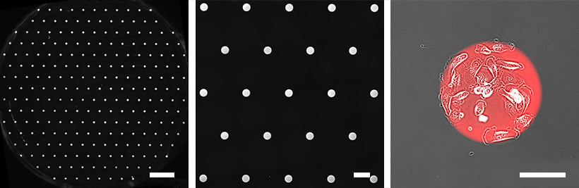
Micropatterning in the µ-Dish 35 mm, high ibiTreat with PLL-PEG passivation created with the ibidi Micro Illumination System. Circular patterns with a diameter of 200 µm and a pitch of 1500 µm, visualized using labeled fibronectin (HiLyte 647, red). Left: Overview of the entire µ-Dish growth area. Middle: Detailed view of the patterned surface. Right: MDA-MB-231 cells seeded onto the patterns show specific adhesion to the defined geometries within 2 hours post-seeding. Refer to Application Note 73 for the complete protocol.
Micropatterning for Spheroid Generation
Spheroid formation of NIH-3T3 cells, seeded on a 200 µm RGD pattern in the channels of the µ-Slide VI 0.4 over a period of 40 hours. Patterns were generated with the ibidi Micro Illumination System. Refer to Application Note 72 for the complete protocol.
abricate Custom Microdevices
Photolithography, soft lithography, and photopolymerization enable the fabrication of defined 2D and 3D microstructures. Photocrosslinkable hydrogels, bioinks, photoresists, and 365 nm photoinitiators can be used to create custom molds, microarchitectures, and structured biomaterials.
Passive Flow Control with Tesla Valve Networks
Channel-in-a-channel structure created with the ibidi Micro Illumination System by using a photo-crosslinkable PEG-norbornene hydrogel and the µ-Slide I 0.4 Luer. The Tesla valve hydrogel channel was visualized by injecting blue food coloring.
PDMS Master Mold Fabrication
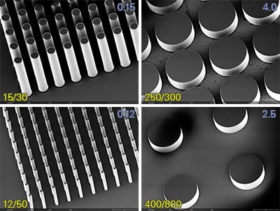
Scanning Electron Microscopy (SEM) image of a 3″ silicon wafer with a structured SU-8 photoresist layer as a PDMS master mold for microscopic structures, created with the ibidi Micro Illumination System. The yellow annotations indicate the diameter and center-to-center distance of these structures, while the blue annotations denote their aspect ratio. Refer to Application Note 75 for the complete protocol.
Specifications
| Light Source | High-power UV LED |
| Wavelength | 365 nm |
| Intensity | 20 mW/cm² (@20%) – 100 mW/cm² (@100%) |
| Time | 1 s – 59 min, 59 s |
| Divergence | ≤ 3° |
| Illumination area | ⌀ 70 mm |
| Compatible photomasks | 3″ masks, thickness 1.4–1.8 mm |
| Nominal resolution | 2 µm |
| Recommended feature size | >10 µm |
| Outer dimensions (l × w × h) | 200 mm × 215 mm × 260 mm |
| Weight | 6.6 kg / 14.6 lbs |
| Material | Multi-material construction |
Technical Features
Inverted UV-Illumination
- Large homogeneous UV-illumination area for sharp pattern transfer due to the highly collimated 365 nm LED
- UV exposure from below the sample, ideal for use with coverslip-bottom labware (e.g., µ-Slides and µ-Dishes)
Compact, Turnkey, and Flexible
- Small footprint benchtop system—no external equipment or infrastructure needed
- Intuitive user-friendly handling—no software installation or computer interface required
- Flexible usage—versatile microstructuring applications
Photomask Compatibility
- Supports standard 3″ photomasks for versatile patterning applications
Integrated Safety Lid
- Equipped with a secure lid and safety lock mechanism