HALO® | Quantitative Image Analysis for Pathology คือแพลตฟอร์มซอฟต์แวร์วิเคราะห์ภาพดิจิทัลสำหรับพยาธิวิทยา (Digital Pathology) จาก Indica Labs ร่วมกับ Leica Biosystems ซึ่งช่วยให้การวิเคราะห์ภาพ Whole‑Slide Image (WSI) เป็นไปอย่างมีประสิทธิภาพ แม่นยำ และขยายผลได้ตามความต้องการของงานวิจัยหรือทางคลินิก
ภาพรวมแพลตฟอร์ม HALO®
- ใช้งานง่าย รวดเร็ว เรียนรู้ได้ทันที
ออกแบบให้ทุกระดับผู้ใช้สามารถเรียนรู้และใช้งานได้ไม่ยุ่งยาก ผ่านอินเทอร์เฟซที่ตรงไปตรงมาและโมดูลที่สร้างไว้แล้วพร้อมใช้ - รองรับภาพทั้ง Brightfield และ Fluorescence
ไม่ว่าจะเป็นสไลด์ H&E, IHC, ISH, FISH หรือ multiplexed panels ก็รองรับได้ทั้งหมด แล้ววิเคราะห์ข้อมูลเชิงตำแหน่งและปริมาณบนเซลล์ได้ละเอียดถึงระดับวินาที - AI‑Ready เพื่องาน image analysis
เลือกใช้โมเดล AI สำเร็จรูปอย่าง nuclear segmentation และ tissue classifier หรือสามารถฝึกAI ด้วยตัวเองผ่าน HALO AI module ด้วยการลากวาด annotation ง่าย ๆ และ tuning แบบเรียลไทม์
| โมดูล | การใช้งานหลัก |
|---|---|
| Multiplex IHC | นับเซลล์แท็กสีต่างกัน จำนวนสูงสุด 5 ตัวบนภาพ brightfield |
| Highplex FL | วิเคราะห์ biomarker นับไม่จำกัดในภาพ fluorescence |
| Tissue Classifier | จำแนกเนื้อเยื่อต่างชนิดด้วยระบบเรียนรู้จากตัวอย่าง |
| Spatial Analysis | วิเคราะห์การกระจายในพื้นที่ เช่น proximity หรือ infiltration |
| ISH / FISH / FISH‑IF | วิเคราะห์ probe และ cell phenotypes บนสไลด์ chromo หรือ fluorescent |
| TMA, Serial Sect. | วิเคราะห์ภาพ tissue microarray และชุดสไลด์ serial ได้แบบ batch |
| Area Quantification / Object Colocalization | วัดพื้นที่การย้อม, อินเทนซิตี้, colocalization ของโครงสร้างต่าง ๆ |
Image Analysis Simplified
With unmatched ease-of-use, scalability, and powerful analytic capabilities, research, pharma and healthcare organizations worldwide are using HALO® from Indica Labs for high-throughput, quantitative tissue analysis. Spend less time learning the software and more time analyzing your research. The intuitive software design enables analysts at all levels to quickly learn and utilize the system. A comprehensive range of purpose-built HALO modules simplify the analysis workflow so researchers do not need to “build” algorithms from scratch.
Extensive Analytics, Reliable Results
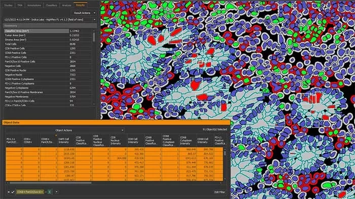
HALO reports morphological and multiplexed expression data on a cell-by-cell basis across entire tissue sections while maintaining an interactive link between cell data and image. Sort and filter cell data to mine millions of cells while also visually assessing cell populations in the context of tissue architecture, leverage interactive markups to understand colocalization and explore combined phenotypes, and define custom analysis outputs.
Leverage Integrated AI Tools
Use pre-trained deep-learning networks for optimized nuclear and membrane segmentation directly in the HALO platform with cell analysis modules in brightfield or fluorescence, including the Multiplex IHC and Highplex FL modules.
Want to train your own AI? Learn more about HALO AI.
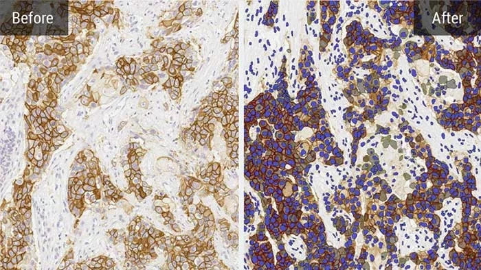
AI Powered Nuclear and Membrane Segmentation
With pre-trained AI-based networks for nuclear and membrane segmentation available in both brightfield and fluorescence, optimizing your segmentation parameters has never been easier.
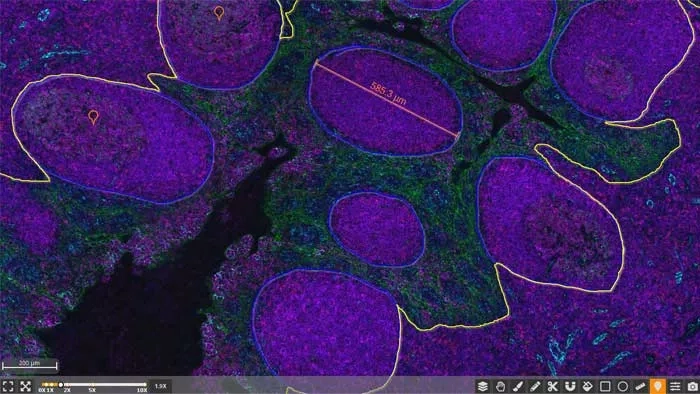
Annotation Tools and Markups
Annotate and explore images with a variety of freehand tools, reticle-based tools, toggle markup images, explore intensity data, measure distances, or take a quick snapshot.
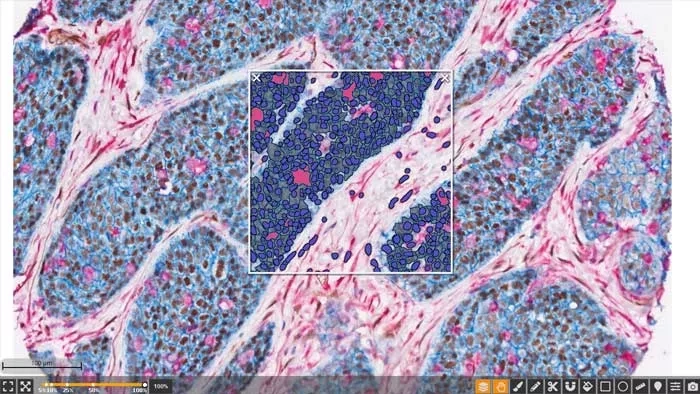
Users can easily toggle Interactive markups on and off to investigate results within each cell population of interest.
Comprehensive Tools
The powerful real-time tuning feature of HALO provides live feedback on image analysis parameters for ease of algorithm optimization. Built-in heatmap functionality provides spatial insights such as immune cell density surrounding a tumor, while image registration allows for serial IHC-stained slide viewing and synchronized navigation.
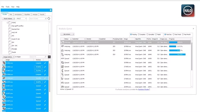
Your Analysis, Your Way
Customize image analysis outputs and set up calculations to deliver the data you need. Use saved analysis settings to queue batch analysis to run silently in the background while you continue to work in HALO.
Advanced data export features ensure you have the output you need, including exporting annotations, analysis or classifier settings, reports, summary data, object data in a spreadsheet or FCS format.
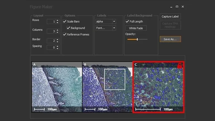
Figure Maker
Acquire publication quality figures in just a few clicks with the figure maker feature. Quickly define the number of frames, select images of interest, then optimize scale bars, labels, fonts, opacity, and more. Save at your desired image size and resolution as a .png, .tif, or .jpg.