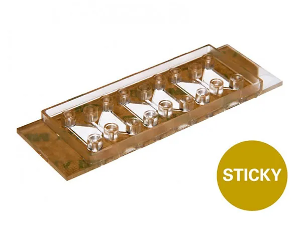
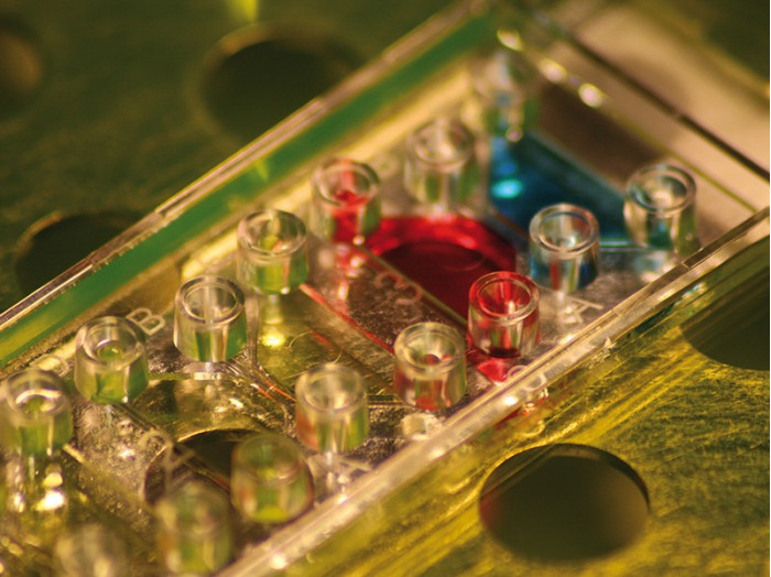
สไลด์แบบ chemotaxis แบบไม่มีก้น โดยด้านล่างมีกาวในตัวซึ่งสามารถติดที่พื้นผิวได้
- อเนกประสงค์ โดยสามารถใช้ติดกับวัสดุที่หลากหลาย
- สำหรับการวัด chemotaxis แบบ real time
- มีความสเถียรสำหรับการทดลองในระยะยาว
A chemotaxis bottomless slide used in 2D and 3D applications with a self-adhesive underside designed to allow self-assemblage of the slide setup
- Versatile – i.e., permits the mounting of a variety of bottom materials
- Allows chemotaxis measurements to be taken in real-time
- Provides stable gradients for long-term experiments
- Pcs./Box: 10 (individually packed)
Applications:
- Inserting materials or tissue into chemotaxis chambers
- Keeping cell spheroids or tissue in a stable concentration gradient
- Chemotaxis of fast or slow migrating cells on either a 2D surface or in 3D gels
- Usable with a range of bottom materials, such as plastic sheets, silicon chips, and glass slides
Specifications:
| Outer dimensions (w x l) | 25.5 x 75.5 mm² |
| Chemotaxis chambers on slide | 3 |
| Volume per chamber | 130 µl |
| Observation area | 2 x 1 mm² |
| Height of reservoirs | 1.05 mm |
| Total height with plugs | 12 mm |
| Volume chemoattractant | 30 µl |
| Bottom | none |
Technical Features:
- Bottomless slide
- Self-adhesive underside
- Biocompatible adhesive that has been cell culture tested
- Adheres to all flat surfaces, even wet surfaces
- Suitable coverslips available
- Specifications identical to those of the µ-Slide Chemotaxis, except that it has no bottom

Placing Tissue Sample in the sticky-Slide Chemotaxis


Application Example: Directed Invasion of MCF-7 Cancer Cells in an FCS Gradient
This movie shows the invasion of MCF-7 cancer cells into a collagen gel during 48 hours. The center channel of a sticky-Slide Chemotaxis was increased in height by milling in order to position the MCF-7 spheroid. The left side contains 10% fetal calf serum, the right side is serum-free. Phase contrast microscopy, objective lens 4x.

Technical Drawing

Technical drawings and details are available in the Instructions (PDF).
