Leica – Visoria P เป็นกล้องจุลทรรศน์โพลาไรซ์ที่ออกแบบมาเพื่อศึกษาสมบัติทางแสงของวัสดุและตัวอย่างทางธรณีวิทยา โดยใช้แสงโพลาไรซ์เพื่อเน้นรายละเอียดที่มองไม่เห็นด้วยแสงปกติ ช่วยเพิ่มประสิทธิภาพในการทำงานด้วยฟังก์ชันเข้ารหัส, ระบบจัดการแสงอัตโนมัติ และการออกแบบตามหลักสรีรศาสตร์ที่ช่วยลดความเมื่อยล้า
Leica – Visoria P เป็นกล้องจุลทรรศน์โพลาไรซ์ที่ออกแบบมาสำหรับการศึกษาวัสดุที่มีคุณสมบัติแสงสองแนว เช่น แร่ หิน ถ่านหิน พลาสติก พอลิเมอร์ ผลึกเหลว แก้ว และคอนกรีต ช่วยให้เห็นรายละเอียดภายในที่ไม่สามารถตรวจสอบได้ด้วยแสงปกติ เหมาะสำหรับงานด้านธรณีวิทยา วิศวกรรมวัสดุ และการวิจัยเชิงแสง
กล้อง Leica – Visoria P ช่วยให้คุณประหยัดเวลาในการทำงานด้วยระบบจัดการแสงอัตโนมัติ ไม่ต้องปรับความสว่างเองเมื่อต้องเปลี่ยนกำลังขยายหรือโหมดคอนทราสต์ ระบบจะจดจำและปรับแสงให้เหมาะสมโดยอัตโนมัติจากการเข้ารหัสของตัวกล้อง ทำให้คุณสามารถโฟกัสกับการวิเคราะห์ตัวอย่างได้ต่อเนื่อง
กล้อง Leica – Visoria P ช่วยให้การบันทึกภาพตัวอย่างเป็นเรื่องง่ายและรวดเร็วด้วยปุ่มถ่ายภาพที่อยู่บนฐานกล้อง พร้อมบันทึกการตั้งค่าระบบและข้อมูลเมตาอัตโนมัติเมื่อจัดเก็บภาพ แถบสเกลจะถูกเพิ่มและปรับขนาดอัตโนมัติเพื่อความแม่นยำและประหยัดเวลา
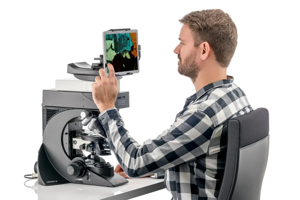
กล้อง Leica – Visoria P รุ่นดิจิทัลแบบไม่มีเลนส์ตา ช่วยให้คุณทำงานได้อย่างสบาย โดยสามารถดูภาพผ่านแท็บเล็ต ลดอาการเมื่อยล้าจากการใช้งานระยะยาว บันทึกและแชร์ภาพได้สะดวก และประหยัดพื้นที่บนโต๊ะทำงานโดยไม่ต้องใช้คอมพิวเตอร์
Leica – Visoria P ออกแบบตามหลักสรีรศาสตร์ ช่วยให้ผู้ใช้ทำงานได้อย่างสบาย ลดอาการเมื่อยคอและหลังจากการใช้งานเป็นเวลานาน ด้วยปุ่มปรับโฟกัสที่สามารถปรับความสูงได้ และตำแหน่งที่จัดวางอย่างสมมาตรเพื่อให้มือและแขนอยู่ในท่าที่เหมาะสม
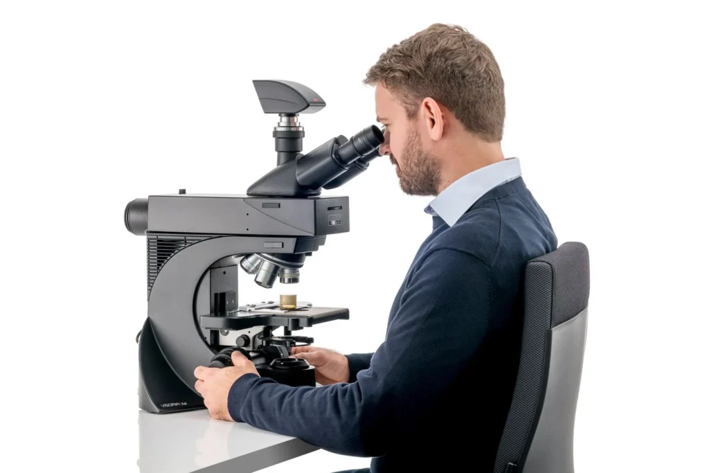
Leica – Visoria P สามารถปรับให้เหมาะกับสรีระผู้ใช้งานได้ ด้วยอุปกรณ์เสริมที่ช่วยให้คุณนั่งทำงานในท่าทางที่ถูกต้อง ลดความเมื่อยล้าระยะยาว เช่น ErgoModules สำหรับปรับความสูงของเลนส์ตา และ ErgoLift ที่ช่วยปรับระดับความสูงของตัวกล้องได้อย่างสะดวก
Conoscopy เป็นเทคนิคที่ใช้ศึกษาสมบัติทางแสงของวัสดุที่มีลักษณะแสงไม่สม่ำเสมอ (anisotropic) เช่น แร่หรือผลึก โดย Leica – Visoria P รองรับการใช้งาน conoscopy ผ่านโมดูลเสริม 3 แบบให้เลือก ได้แก่:
- Bertrand lens cube
- Bertrand lens module (A/B)
- Advanced conoscopy module
แต่ละโมดูลให้ระดับความสามารถที่ต่างกัน ขึ้นอยู่กับความต้องการของงานวิเคราะห์
Leica – Visoria P รองรับการวิเคราะห์ถ่านหินและแร่ใยหิน (asbestos) ด้วยเลนส์วัตถุเฉพาะทาง:
- สำหรับถ่านหิน: ใช้เลนส์ immersion ชนิดน้ำมันเพื่อช่วยแยกแยะองค์ประกอบต่าง ๆ ของตัวอย่างได้แม่นยำขึ้น
- สำหรับใยหิน: ใช้เลนส์วัตถุชนิดพิเศษสำหรับเทคนิค dispersion staining ซึ่งช่วยให้เห็นการกระจายสีที่บ่งชี้ลักษณะเฉพาะของใยหิน
กล้อง Leica – Visoria P ทำงานร่วมกับซอฟต์แวร์ Enersight เพื่อเพิ่มประสิทธิภาพในการสังเกตและวิเคราะห์ตัวอย่าง ใช้งานง่ายผ่านอินเทอร์เฟซเดียว ช่วยให้ได้ภาพความละเอียดสูง มุมมองกว้าง ปรับแสงอัตโนมัติ และรวมภาพจากหลายเทคนิคคอนทราสต์ เช่น brightfield และ polarization เพื่อให้เข้าใจโครงสร้างวัสดุได้ลึกยิ่งขึ้น
Visoria P Polarization Microscope
Power up routines with efficiency and comfort
| Microscope | |
| Size and weight | Length: 410 mm, width: 331 mm, height: 505 mm, approx. 18 kg (depending on configuration) |
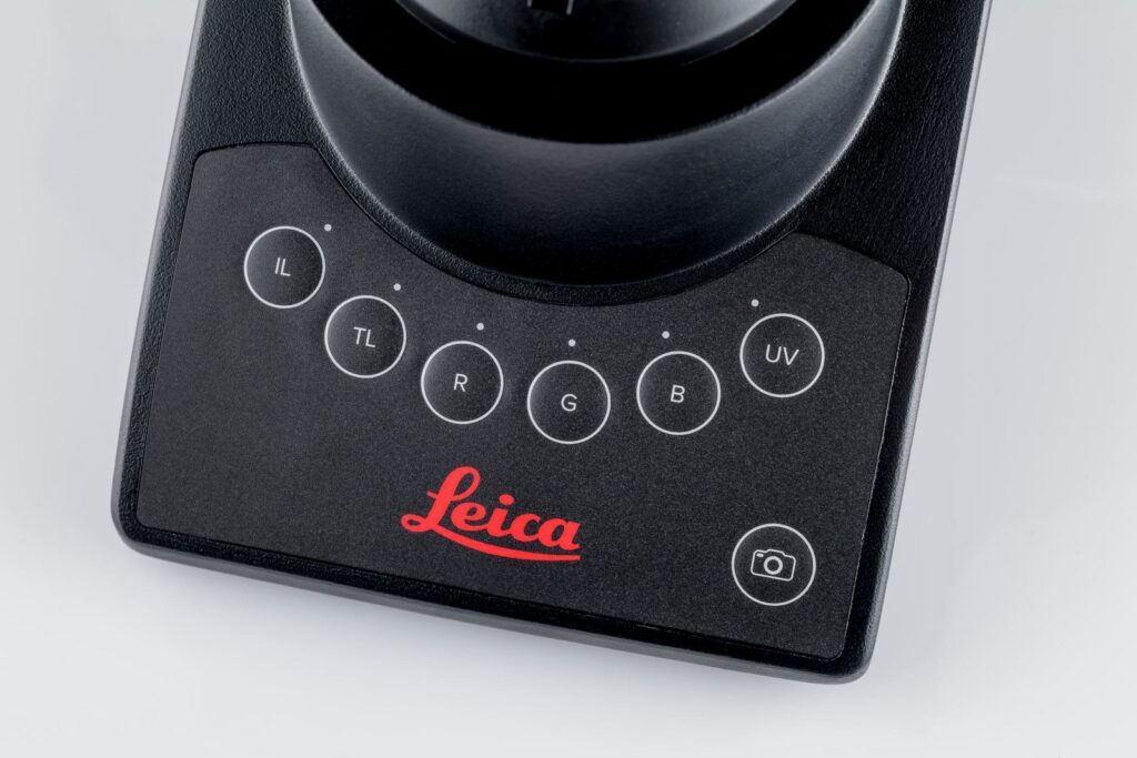
| Stand | Illumination toggle buttons with status indicator, image capture button, antimicrobial surface with AgTreat (ISO 22196) |
| Optics | |
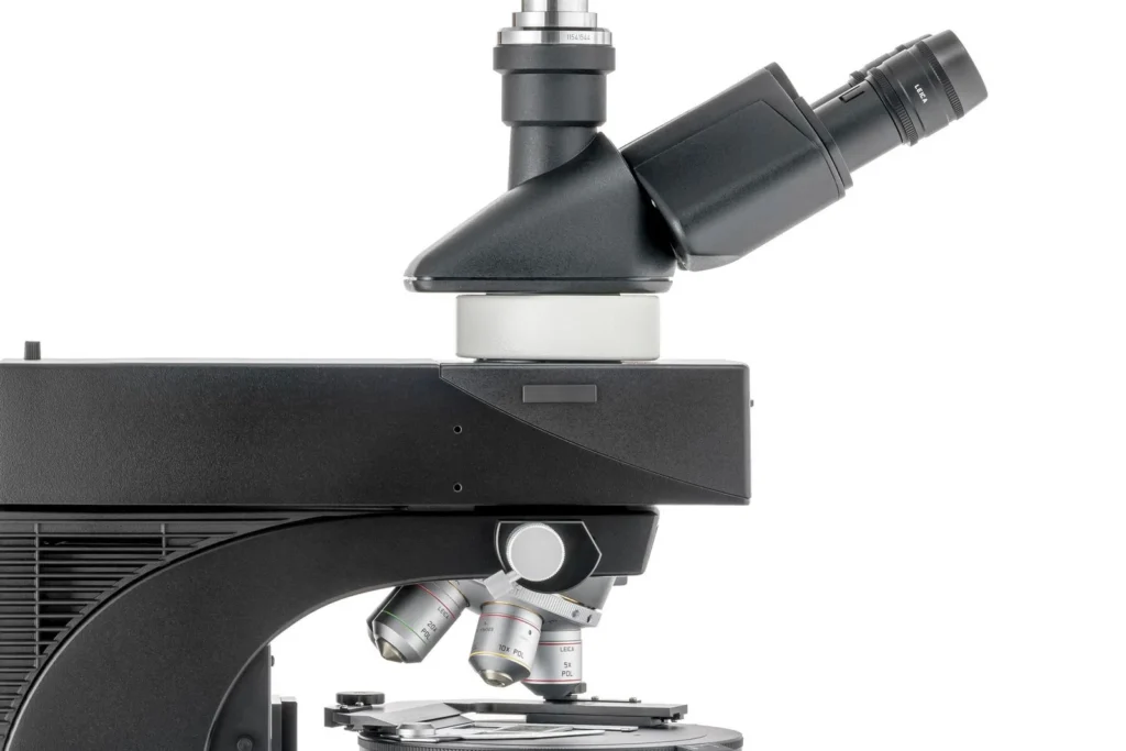
| Nosepiece | Encoded 5x (M25), centerable |
| Eyepieces (FOV) | 20 / 22 / 25 mm |
| Tubes | Phototubes 50 / 50 with fixed port, 100 / 50 / 0 with fixed port, 100 / 50 / 0 with variable port |
| Digital version | Digital version with 10” screen / tablet |
| Ergonomic accessories | Wide range of ergonomic accessories available (ErgoTubes, ErgoLift, ErgoModules) |
| Encoded illumination management | IL and TL: High-power white LED, encoded 4-color fluorescence illumination, further external light sources on request (non-encoded) |
| Incident light axis | Manual encoded, 4-fold filter turret, color-coded diaphragm assistant; field and aperture diaphragm, slots for analyzer / polarizer, two filter positions |
| Fluorescence light axis | Optional |
| Incident light (IL) | Methods: Brightfield (with BF cube or Smith reflector), DIC, fluorescence, oblique illumination, qualitative and quantitative polarization |
| Transmitted light axis | Manual, fixed and flip-top condenser operation with color-coded diaphragm assistant |
| Transmitted light (TL) | Methods: Brightfield, darkfield, phase contrast, DIC, qualitative and quantitative polarization |
| Operation | 360° rotatable polarization stage with verniers and brake, 360° rotatable polarization stage with verniers, 45° clickstop position and brake. Stages are exchangeable and height adjustable. Further stages on request. |
| Stage | Scanning stage 75 x 50 mm, additional manual xy stages available, stage bracket for adaptation of 3rd party stages, bject clamps, sample holder without point counting, sample holder with point counting |
| Focus drive | Height-adjustable focus knobs, 19 mm travel range, maximum 28 mm total stage stroke depending on stage and condenser type, 2-gear focus drive (coarse / fine) with 1 mm scale, 3-gear focus drive (coarse / medium / fine) with 140, 4 and 1 µm scale, torque adjustment, and adjustable upper focus stop |
| Accessories | |
| Conoscopy | Bertrand lens cube, Bertrand lens module (A/B module), advanced conoscopy module (focusable) |
| Analyzer | Fixed, 180°, 360° |
| Polarizer | Fixed, 0 / 45 / 90°, 90° with rotatable lambda plate, 360°, fixed with lambda plate |
| Compensators | Tilting compensators up to 5 or 30 orders, quartz wedge, lambda and 1/4 lambda plate |
| General specifications | |
| Supply voltage | 100–240 V AC, 50 / 60 Hz, power consumption max. 15 W |
| Ambient conditions | 15–35°C, relative humidity max. 80% up to 30°C (non-condensing) |