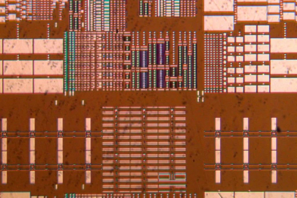
Leica – Visoria M คือกล้องจุลทรรศน์สำหรับการตรวจสอบวัสดุในงานทั่วไป เช่น โลหะ โลหะผสม ชิ้นส่วนอิเล็กทรอนิกส์ กลไก เซรามิก และวัสดุผสมต่าง ๆ เมื่อนำมาใช้ร่วมกับซอฟต์แวร์ Enersight ยังสามารถวิเคราะห์โครงสร้างหน้าตัดและความหนาของชั้นวัสดุได้อย่างแม่นยำ
Leica – Visoria M ช่วยให้คุณประหยัดเวลาในการทำงานด้วยระบบจัดการแสงอัตโนมัติ ไม่ต้องปรับความสว่างเองเมื่อต้องเปลี่ยนกำลังขยายหรือโหมดคอนทราสต์ ระบบจะปรับแสงให้เหมาะสมโดยอิงจากการตั้งค่าที่เข้ารหัสไว้ในตัวกล้อง ทำให้คุณสามารถโฟกัสกับการสังเกตตัวอย่างได้ต่อเนื่องและแม่นยำยิ่งขึ้น
กล้อง Leica – Visoria M ช่วยให้การบันทึกภาพตัวอย่างเป็นเรื่องง่าย ด้วยปุ่มถ่ายภาพที่เข้าถึงสะดวก สามารถกดบันทึกได้ทันทีโดยไม่ต้องละสายตาจากภาพ ระบบจะจัดเก็บค่าการตั้งค่าพร้อมข้อมูลเมตาโดยอัตโนมัติ และเพิ่มแถบสเกลปรับขนาดอัตโนมัติเพื่อประหยัดเวลาและเพิ่มประสิทธิภาพในการทำงาน
กล้อง Leica – Visoria M ถูกออกแบบมาให้ใช้งานได้อย่างง่ายดาย เหมาะสำหรับงานประจำที่ต้องการความรวดเร็วและแม่นยำ มีระบบรหัสสีช่วยเลือกช่องแสงให้เหมาะกับเลนส์แต่ละตัว, ระบบหยุดโฟกัสในตัวช่วยป้องกันความเสียหายต่อตัวอย่าง และมีปุ่มโฟกัสแบบ 3 ระดับเพื่อการปรับภาพที่แม่นยำในทุกระดับการขยาย
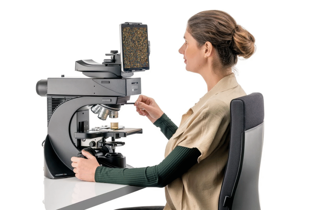
กล้อง Leica – Visoria M รุ่นดิจิทัลไม่มีเลนส์ตา ช่วยให้ผู้ใช้งานสามารถดูภาพผ่านแท็บเล็ตได้โดยตรง ลดอาการเมื่อยล้า ใช้งานง่าย ประหยัดพื้นที่ ไม่ต้องใช้คอมพิวเตอร์ และสามารถบันทึกหรือแบ่งปันข้อมูลกับเพื่อนร่วมงานได้อย่างสะดวก
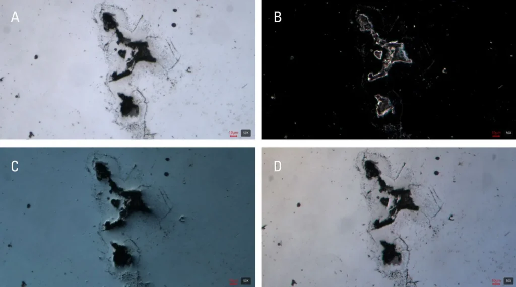
Visoria M ช่วยให้คุณมองเห็นรายละเอียดของโครงสร้างและตำหนิต่าง ๆ บนตัวอย่างได้ชัดเจน ด้วยเทคนิคคอนทราสต์หลากหลาย เช่น brightfield, darkfield, polarization, DIC, fluorescence และเฉพาะทางอย่าง oblique illumination ที่เหมาะสำหรับการดูพื้นผิวที่มีมิติสูง เช่น รอยขีดข่วนหรือการปนเปื้อนบนวัสดุ
เลนส์เสริม 0.7x Macro ของ Leica – Visoria M ช่วยให้มองภาพรวมของตัวอย่างได้รวดเร็วในมุมมองกว้างประมาณ 36 มม. เหมาะสำหรับการสแกนหาตำแหน่งหรือจุดสนใจเบื้องต้น ก่อนจะซูมดูรายละเอียดด้วยเลนส์ขยายที่สูงขึ้น ช่วยประหยัดเวลาเมื่อเทียบกับเลนส์ทั่วไป
กล้อง Visoria M ทำงานร่วมกับแพลตฟอร์มซอฟต์แวร์ Enersight เพื่อช่วยให้การวิเคราะห์ตัวอย่างวัสดุมีประสิทธิภาพมากขึ้น ไม่ว่าจะเป็นการวัดความหนาชั้นเคลือบ การรวมภาพเพื่อดูพื้นที่กว้าง (XY Stitching), การได้ภาพคมชัดลึก (EDOF), ปรับแสงอัตโนมัติ และการรวมภาพจากหลายเทคนิคคอนทราสต์ เช่น brightfield และ darkfield เพื่อให้เห็นรายละเอียดตัวอย่างชัดเจนยิ่งขึ้น
Visoria M Materials Microscope
Power up routines with efficiency and comfort
| Microscope | |
| Size and weight | Length: 410 mm, width: 331 mm, height: 505 mm, approx. 18 kg (depending on configuration) |
| Stand | Illumination toggle buttons with status indicator, image capture button, built-in analyzer slot, antimicrobial surface with AgTreat according to ISO 22196 |
| Optics | |
| Nosepiece | Encoded 5x (M32), encoded 6x (M25) |
| Eyepieces (FOV) | 20 / 22 / 25 mm |
| Tubes | Wide range of standard, ergonomic and phototubes available, with different beam splitters available |
| Digital version | Digital version with 10” screen / tablet |
| Ergonomic accessories | Wide range of ergonomic accessories available (ErgoTubes, ErgoLift, ErgoModules) |
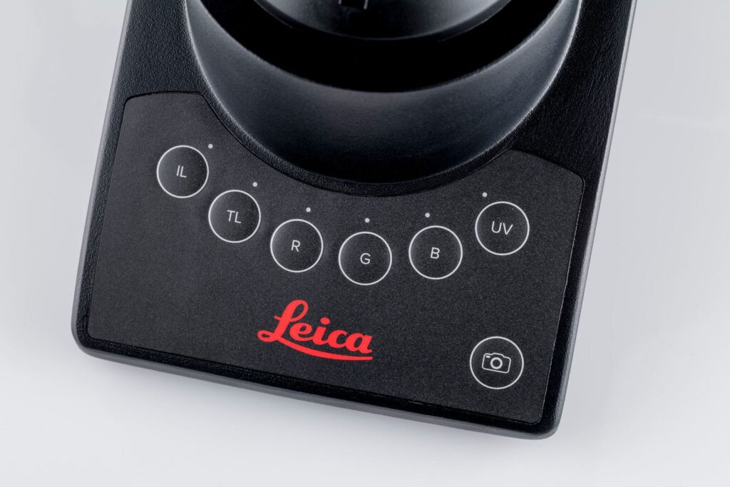
| Encoded illumination management | IL and TL: High-power white LED, encoded 4-color fluorescence illumination, further external light sources on request (non-encoded) |
| Incident light axis | Manual encoded, 4-fold filter turret, color-coded diaphragm assistant; aperture diaphragm, slots for analyzer / polarizer, two filter positions |
| Fluorescence light axis | Optional |
| Incident light (IL) | Methods: Brightfield (with BF cube or Smith reflector), darkfield, DIC, fluorescence, oblique illumination, qualitative polarization |
| Transmitted light axis | Manual, fixed and flip-top condenser operation with color-coded diaphragm assistant |
| Transmitted light (TL) | Methods: Brightfield, darkfield, phase contrast, DIC, qualitative polarization |
| Operation | |
| Stage | Stages are exchangeable and height-adjustable. manual XY-stage 76 x 50 mm / 3-plate stage (4 x 4), additional stages (incl. rotating or large-sample stages) |
| Stage control | Left-, right-handed stage, torque-adjustable handle |
| Focus drive | Height-adjustable focus knobs, 19 mm travel range, maximum 28 mm total stage stroke depending on stage and condenser type, 2-gear focus drive (coarse / fine) with 140, 4 and 1 µm scale, torque adjustment, and adjustable upper focus stop |
| Accessories | |
| Analyzer | Fixed, 180°, 360° |
| Polarizer | Fixed, 0 / 45 / 90°, 90° with rotatable lambda plate, 360°, fixed with lambda plate |
| General specifications | |
| Supply voltage | 100–240 V AC, 50 / 60 Hz, power consumption max. 15 W |
| Ambient conditions | 15–35°C, relative humidity max. 80% up to 30°C (non-condensing) |