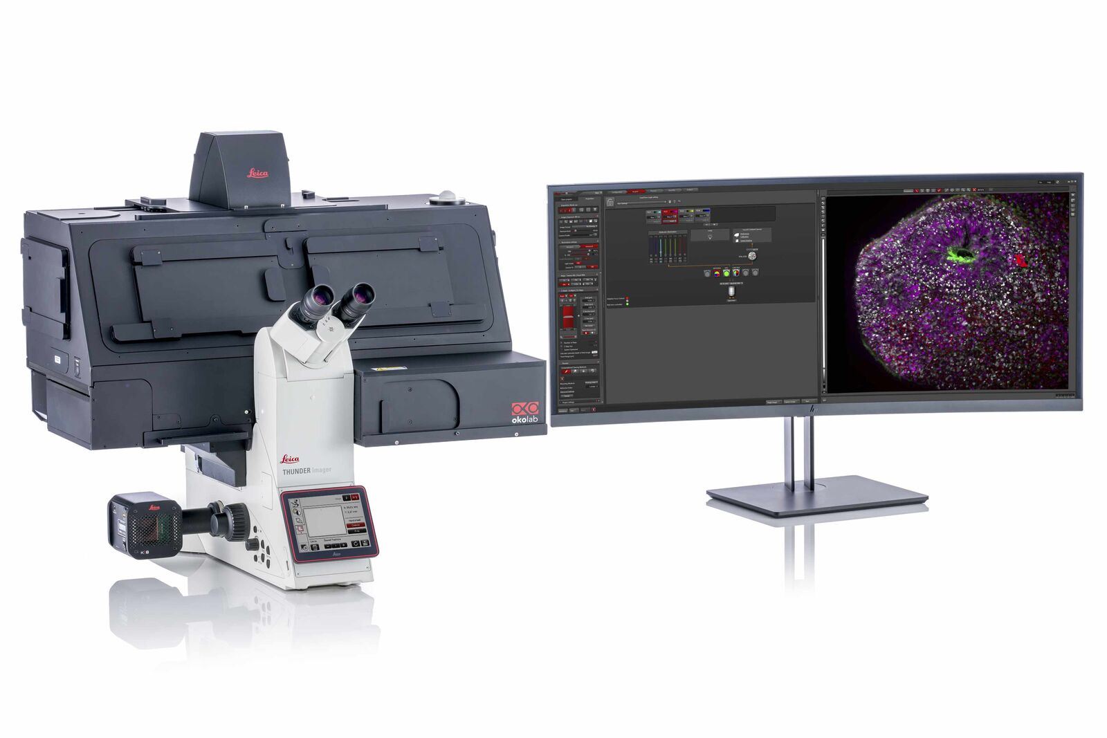
ชุดกล้องจุลทรรศน์หัวกลับ แบบใช้แสงฟลูออเรสเซนต์ พร้อมโปรแกรมประมวลผลแบบ AI ความเร็วสูง ยีห้อ ไลก้า รุ่น Thunder Imager Live Cell และ 3D Assay
โดย Leica THUNDER Imagers ออกแบบมาสำหรับการถ่ายภาพตัวอย่างฟลูออเรสเซนต์แบบ 3 มิติขั้นสูง ไม่ว่าคุณจะต้องการศึกษาสเต็มเซลล์, สเฟียรอยด์ หรือ ออร์แกนอยด์
Leica THUNDER Imagers นำเสนอเทคโนโลยี “Computational Clearing” ซึ่งเป็นนวัตกรรมในการกำจัดความเบลอที่ไม่อยู่ในโฟกัสได้อย่างมีประสิทธิภาพในเวลาจริง ส่งผลให้สามารถถ่ายภาพตัวอย่างเป็นแบบ 3 มิติด้วยกล้องจุลทรรศน์ฟลูออเรสเซนซ์ได้อย่างมีประสิทธิภาพ โดยการทำงานของกล้องมีความไวสูงทำให้มั่นใจได้ว่าตัวอย่างโดยแสงน้อย และไม่เกิด Phototoxicity และ photobleaching
Optimal physiological conditions – minimum light exposure
For 3D cell culture, observing the true physiology is paramount for meaningful results. Typically, you want to investigate your cells while they are in a close to natural state by optimizing experimental conditions, e.g., the lowest light intensity and shortest exposure times possible.
THUNDER Imager Live Cell meets these demands with a high-end LED source that has a small bandwidth optimized for excitation. Even with low illumination and short exposure times, the sensitive high-end sCMOS camera delivers meaningful image information thanks to a quantum efficiency of up to 82%.
To further reduce exposure of the sample to light, illumination is limited to the actual recording time. The camera shutter is synchronized with the high-speed (20 µs switching time) Lumencor LED light source to minimize photobleaching.
Find the THUNDER Imager that’s right for you
Whether you are looking for a dedicated high-end imaging system that excels in a given application, or a versatile solution for a lab running different kinds of assays with various samples, we’ve got you covered.
Here are some selected application examples that show you the strengths of THUNDER:
Master time-lapse multi-position experiments: tracking cell changes
THUNDER Imager Live Cell offer both speed and reliability for your 3D cell culture multiwell experiments. For example, when tracking the growth and development of spheroids and organoids, its speed and reliability allows you to obtain excellent results.
Accurate time-lapse multi-position experiments and tracking of cell changes are achieved with this THUNDER Imager due to:
- Reliable drift correction with the Adaptive Focus Control (AFC)
- Software autofocus that compensates for changes in specimen position
- Reproducible Z-positioning with a precision of up to 20 nm (closed-loop focus)
Get more data points in less time with the new Quantum Stage. It moves rapidly and accurately (e.g. 10 positions per second) to all positions with an excellent repeatability (< ±0.25 µm), as there is no jiggling.
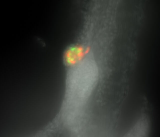
Developing Zebrafish Pancreas
The THUNDER Imager 3D Assay enabled clear identification of Alpha (GFP-green) and Beta (mCardinal-red) cells within a developing zebrafish pancreas.
Imaged in the blue (Hoechst), green (GFP), and red (mCardinal) channels, this 150-image z-stack was completed with all channels within a minute.
Physiological conditions can be maintained within a sample by minimizing photobleaching, providing high-performance imaging, and high-throughput of data, leading to better workflow efficiency.
 Developing zebrafish pancreas – Widefield imageAfter Computational Clearing – THUNDER Imager 3D Assay

Routine, reliable data acquisition
THUNDER Imager Live Cell enable reliable image data acquisition with accurate focus maintained on the live cells at all times.
Live cell imaging is often tricky, because of drift, morphology change, or growth of the cells. Drift is caused by vibrations, mechanical creep, or temperature fluctuations. Both drift and cell changes can reduce the reliability of the acquired image data, as focus becomes an issue. Thanks to the Adaptive Focus Control (AFC), closed-loop focus, and software autofocus, THUNDER Imager Live Cell reliably maintains focus for your multi well experiments.
Clarity and speed for sensitive sample imaging
Combine THUNDER’s out-of-focus blur removal with the advantages of TIRF. For dynamic processes at the cell surface, Total Internal Fluorescence Microscopy provides outstanding signal-to-background separation.
The two videos here show ins-1 cells expressing human pro-insulin with GFP-GRINCH. As KCL is added to the cell culture, the insulin-producing cells depolarize, and insulin residues can be seen fusing to the plasma membrane.
With the THUNDER Imager Live Cell combined with TIRF, image your sensitive samples with outstanding clarity, speed, and imaging parameter control.
More information on the Infinity TIRF
Image at the speed of life – Cellular processes
Life is fast, especially for a cell. Most modern live cell imaging experiments demand high-speed systems.
THUNDER Imager Live Cell provides you with the power of highly sensitive sCMOS camera-based fluorescence systems working with full frames in one shot.
In combination with high sensitivity, THUNDER Imager Live Cell allows you to capture data at up to 90 frames per second, helping you to observe fast cellular events. It captures blur-free image data quickly even deep inside a thick 3D cell cluster. You can stay on top of fast processes even during multiple emission wavelength experiments thanks to fast-switching external filter wheels (< 27 ms).
The following configurations were used in the applications provided in the examples.
THUNDER Imagers can be configured to your application. Get in contact with us.
| THUNDER Imager | |||
| 3D Assay | Live Cell | Live Cell | |
| Specimen | Retina | Brain organoid | Explant aorta |
| Imaging Method | THUNDER | THUNDER | THUNDER |
| Workflow | Navigator spiral scan | Navigator large tile scan | Long-term live cell culture |
| Stand | DMi8 | DMi8 | DMi8 |
| Focus | Z-Focus, motorized, closed-loop | THUNDER AFC (hardware focus for fast focusing and drift correction) | Motorized, closed-loop, AFC (hardware focus for fast focusing and drift correction) |
| Stage | Scanning stage | QUANTUM high speed stage (real-time controlled with Synapse) | QUANTUM high speed stage (real-time controlled with Synapse) |
| Transmitted light contrast & method | BF, DIC | BF | BF |
| EFW | – | + | + |
| Filter cubes | Single band pass | Quad | Quad |
| Fluorescence illumination | LED3 | LED5 | LED8 |
| Incubation | – | Stage-top incubation | Incubator i8, Temp, CO2 control |
| Detection | K5 | DFC9000 GTC | DFC9000 GTC, Hamamatsu Flash4 |
| Optional Upgrade path for advanced imaging workflows | – | + | + |
| Optional Photomanipulation | – | Infinity Scanner | Infinity Scanner |
| Optional TIRF | – | TIRF | TIRF |
Core strengths of our THUNDER Imagers:
| THUNDER Imager | |||
| Applications | 3D Assay | Live Cell | |
| Monocell layers | ++ | +++ | |
| Organoids or Spheroids | ++ | +++ | |
| Zebrafish or C.Elegans, and other model organisms | ++ | +++ | |
| Whole organs i.e. bio-printed or uncleared tissue of Mos | ++ | +++ | |
| Thick tissue sections, i.e. biopsies or vibratome sections | ++ | +++ | |
| Carriers | |||
| Multi-well plates | +++ | +++ | |
| Chamber slides | ++ | +++ | |
| Glass slides | ++ | +++ | |
| Petri dishes | + | +++ | |
| Techniques | |||
| Live cell imaging (Time lapse) | + | +++ | |
| Contrast Methods (PhaCo, DIC, IMC) | +++ | ++ | |
| TIRF | – | +++ | |
| Photomanipulation | – | +++ | |
| Ablation, i.e. FRAP/FRET | – | +++ | |