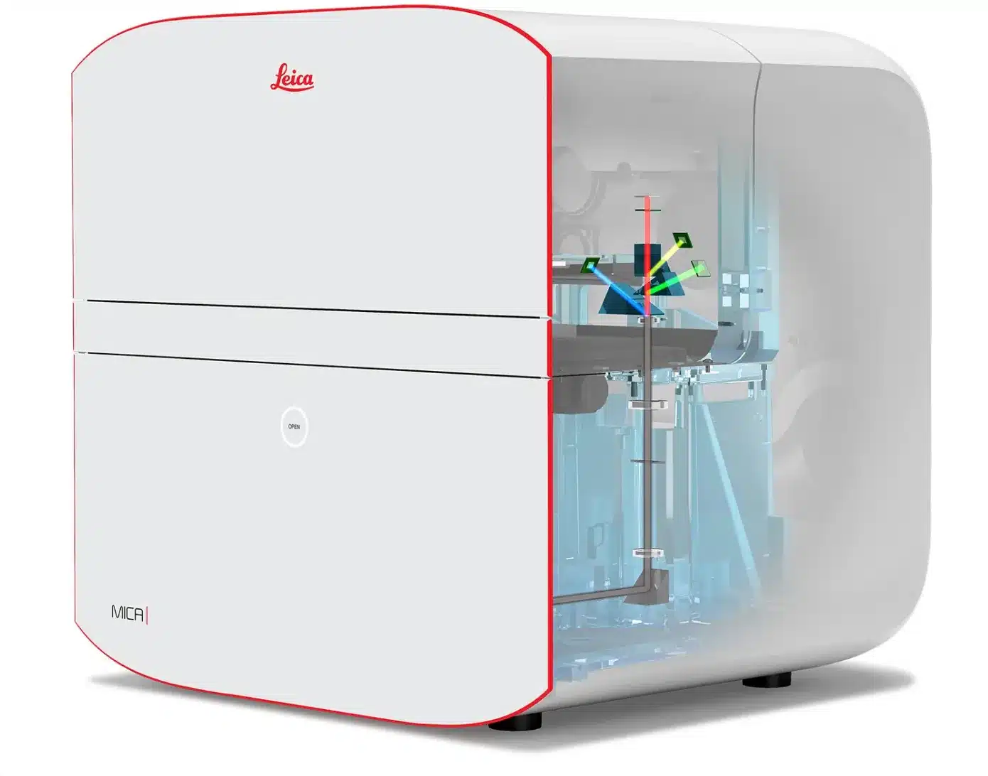
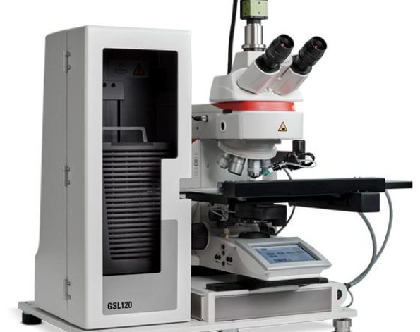
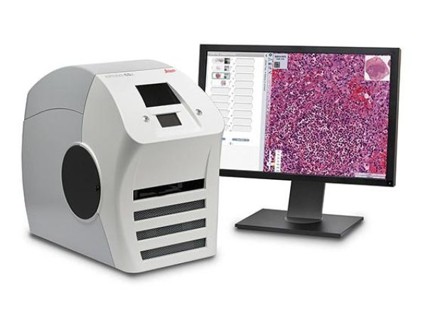
Product : Leica Mica – The world’s First imaging Microhub กล้องจุลทรรศน์ ยี่ห้อ ไลก้า ประเทศเยอรมัน
Type : Confocal Microscope / Advanced Imaging Systems / Fluorescence Microscope / Thunder imager กล้องจุลทรรศน์ชนิดคอนโฟคอล ความละเอียดสูง, กล้องจุลทรรศน์แบบฟลูออเรสเซนต์ / กล้องจุลทรรศน์ชนิด Thunder แบบใช้ปัญญาประดิษฐ์ช่วยวิเคราะห์ภาพ
Model : Mica unites wide field and confocal imaging
Leica Mica เป็นมากกว่ากล้องจุลทรรศน์อัตโนมัติขั้นสูง Mica ได้รวมเทคโนโลยีการถ่ายภาพแบบ Widefield และแบบ Confocal ไว้ในสภาพแวดล้อมที่ถูกปกป้องเป็นพิเศษเพื่อตัวอย่างที่ต้องการบ่มเพาะ
ด้วยการกดเพียงป่มเดียว คุณจะได้ผลลัพธ์ทุกอย่างที่เกี่ยวกับ workflow งานถ่ายภาพเทคนิคฟลูออเรสเซนต์ อย่างรวดเร็ว ทำให้คุณสามารถโฟกัสกับการทดลองทางวิทยาศาสตร์ และไม่ต้องปวดหัวกับการตั้งค่ากล้องจุลทรรศน์อีกต่อไป Mica ช่วยลดข้อจำกัดต่างๆทำให้ขั้นตอนการถ่ายภาพเรียบง่ายอย่างเห็นได้ชัด
- Leica – Mica ทำให้คุณลดขั้นตอนการถ่ายภาพรูปแรกมากถึง 85% และ
- Leica – Mica ช่วยลดเวลาในการถ่ายภาพรูปแรกสูงถึง 33% และ
- Leica – Mica ลดเวลาในการสอนการใช้งานเครื่องได้มากถึง 50% เลยทีเดียว
Leica Mica ช่วยให้คุณได้ข้อมูลเพิ่มขึ้น 4 เท่าตัว โดย Leica Mica – Microhub ช่วยให้คุณสามารถถ่ายภาพตัวอย่างที่ติด 4 แถบ label ที่มีโครงสร้างต่างกัน ได้ในการถ่ายรูปเพียงครั้งเดียว ไม่ว่าจะเป็นการใช้งานในโหมด Widefield หรือ Confocal ก็ตาม โดยไม่ต้องขยับตัวอย่างแม้แต่ครั้งเดียว ด้วยสิ่งนี้ทำให้ตัดปัญหาความคลาดเคลื่อนเชิงพื้นที่ระหว่าง label ของตัวอย่างที่เคลื่อนไหวในขณะถ่ายภาพ ทั้งหมดนี้ได้จากสิทธิบัตรเทคโนโลยีจาก Leica FluoSync ทำให้การถ่ายภาพ Multicolor fluoresence รวมเร็งและสะดวกสบายขึ้น
Leica Mica ช่วยตัดปัญหาการเลือกรูปแบบการถ่ายภาพที่เหมาะสมสำหรับตัวอย่างแบบ Real time
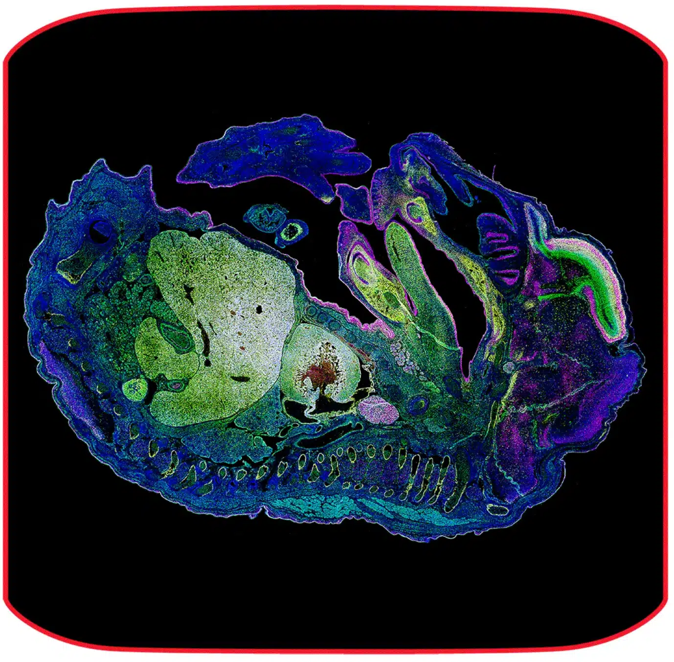
Leica Mica ได้รวมการถ่ายแบบ transmitted, การถ่ายแบบ fluorescence ไว้ในเครื่องเดียวกัน ทำให้คุณสามารถเลือกรูปแบบการถ่ายภาพและเทคนิคต่างๆ เช่น widefield, confocal, THUNDER imaging, LIGHTING, Z-stack, Time-lapse และอื่นๆ อีกมากมายในเครื่องเดียว ด้วยสิ่งนี้ช่วยปลดล็อค ในการสร้างภาพแบบ Overview ที่กำลังขยายต่ำ จนถึงการซูมภาพไปในบริเวณที่สนใจ (Regions of interest) หลังจากนั้นเปลี่ยนโหมดการถ่ายภาพเป็น Confocal ในบริเวณที่ต้องการโดยไม่ต้องย้ายตัวอย่างไปยังระบบอื่นอีกต่อไป
Leica Mica ทำให้ลบข้อจำกัดในการทดลองภายใต้เงื่อนไขของสรีรวิทยา เนื่องจากการทดลองต้องการใช้เซลล์เติบโตในรูปทรงที่เหมาะสมที่สุด โดยทั่วไปแล้ว เซลล์แบบ 2 มิติ และเซลล์แบบ 3 มิติ ในตัวอย่างจะต้องถูกควบคุมในสภาวะแวดล้อมของ อุณหภูมิ และ pH ผ่านทาง CO2 ที่เหมาะสม ดังนั้นความเข้มข้นของสารอาหารและไอออนที่คงที่ต้องการการระเหยที่น้อยที่สุด และมากกว่านั้นบางการทดลองต้องควบคุมความต้องการของก๊าซออกซิเจนด้วย เพื่อเลียนแบบรูปแบบทางสรีรวิทยาของตัวอย่างให้เหมาะสมที่สุด ดังนั้น Leica Mica ช่วยให้การเตรียมเงื่อนไขในการทดลองนี้ให้เหมาะสมที่สุดได้ อีกทั้งยังสามารถควบคุมความมืดและความสว่างสำหรับการทดลองเพื่อเฝ้าดูการทดลองของคุณได้ตลอดเวลา
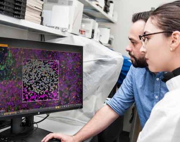
Leica Mica มีะรบบอัตโนมัติอัจฉริยะ และระบบ AI ปัญญาประดิษฐ์ที่ช่วยสนับสนุนการวิเคราะห์ข้อมูลให้มีประสิทธิภาพ ทำให้ท่านสามารถตีพิมพ์ผลงานได้อย่างรวดเร็ว Leica Mica ช่วยลดข้นตอนกระบวนการมากกว่า 60% ผ่านระบบอัจฉริยะ และช่วยทำให้การทดลองซ้ำของคุณได้ผลแม่นยำถึง 100%
0:00
/ 1:02
Access for all
Everyone can now leverage microscopy to make more discoveries.
Mica provides a clear sample overview and allows to easily change observation conditions with just a few clicks.
- 85% fewer steps to the first image
- 33% less time to the first image
- 50% of the training time
No constraints – 4x more data with 100% correlation
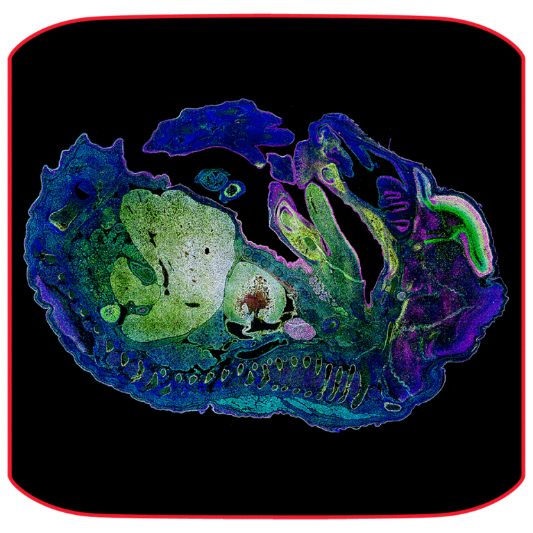
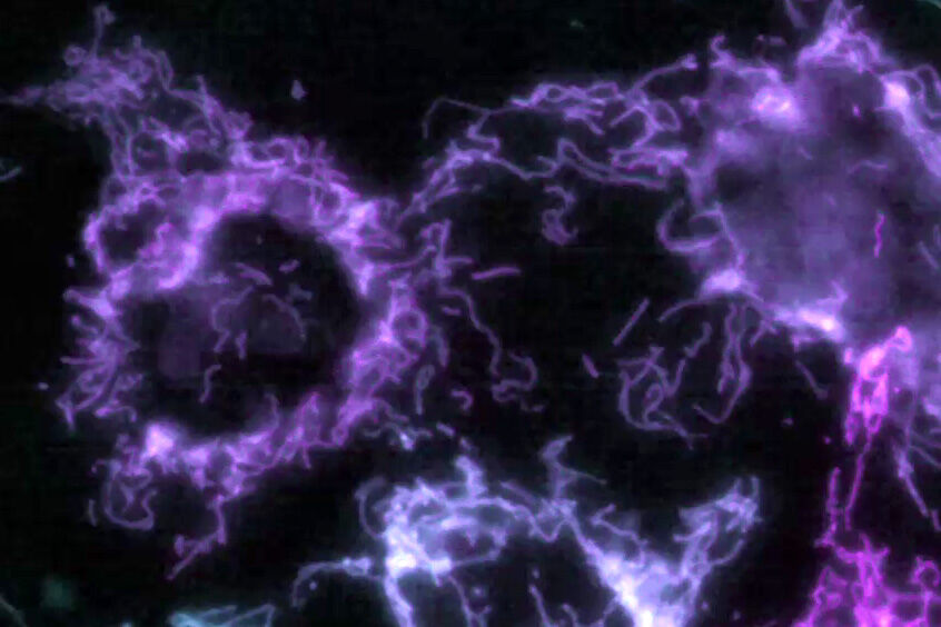
The Microhub enables you to simultaneously capture all 4 labels of different structures in a single acquisition for widefield or confocal, without ever moving your sample. This overcomes the spatiotemporal mismatch between labels of moving objects during sequential acquisition. All powered by our patented FluoSync technology, a fast and gentle method for multicolor fluorescence imaging.
No constraints – Select the right modality in real time
Mica unifies transmitted and fluorescence light imaging modalities. You can select from multiple imaging modalities all within one Microhub, including widefield, confocal, THUNDER imaging, LIGHTNING, Z-stacks, time-lapse and more.
This enables you to
- generate fast overviews with widefield at low magnification
- gradually zoom in on the regions of interest
- switch to confocal when and where needed without ever moving the sample to a different system
No constraints – Achieve physiological-like conditions throughout your experiments
Live cell experiments require the cells to be in optimal shape. Typically, 2D and 3D cells in media require the temperature and the pH (via CO2) in the environment to be controlled. Stable nutrition and ion concentrations require evaporation to be minimal. Some experiments even demand the O2 to be mimicked closer to physiological levels. Mica can provide the right conditions in the live cell configuration.
- Mica is an incubator: the entire encapsulated inner sample space can be climate controlled (temperature, CO2 and humidity regulation) and offers ideal conditions for short and long-term live cell observation.
- From dark to light: Mica also enables you to enjoy a brightly lit lab—freeing you from the constraints of sitting in a dark room for hours monitoring your experiment.
Radically simplified workflows
Intelligent automation and AI-supported analysis enables greater efficiency and a faster track to publication.
- Reduce over 60% of process steps through system intelligence
- Reduce time and effort from sample to insight by simplifying your entire workflow
- Enable 100% reproducibility and repeatability throughout your experiment
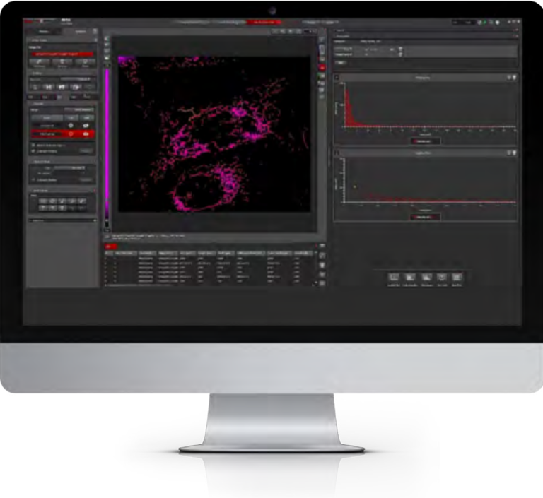
Meet Mica in key applications
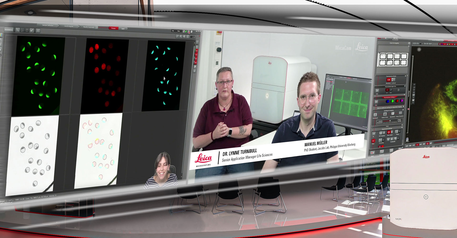
MicaCam Experiments
MicaCam is where life science researchers come together to interact and make discoveries.
Watch relevant scientific experiments using Mica, the world’s first Microhub.
The Microhub era is now!
Experience the future.
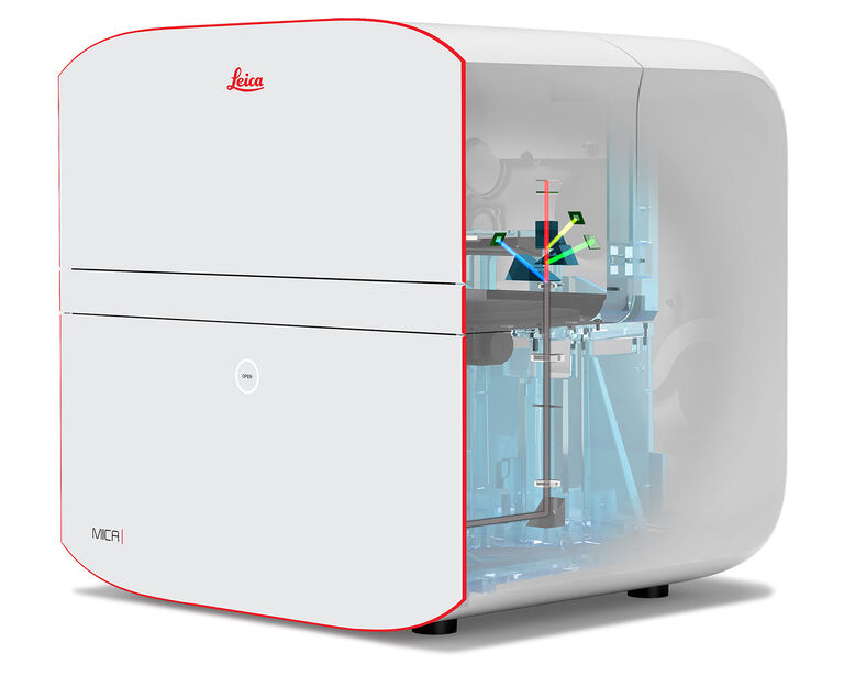
Technical Specifications
| Mica Widefield | Mica Widefield Live Cell | Mica WideFocal | Mica WideFocal Live Cell | ||
| Transmitted Light Contrast | |||||
| Integrated modulation contrast (IMC), automatically adjusted and Brightfield contrast in RGB or gray scale mode | x | x | x | x | |
| Incident Fluorescence Illumination | |||||
| LED | 365 nm, 470 nm, 555 nm, 625 nm | x | x | x | x |
| FluoSync Widefield Detection | |||||
| Simultaneous Detection channels | 4 with FluoSyncTM fluorophore separation | x | x | x | x |
| Detector type | 5 MP CMOS | x | x | x | x |
| Confocal Illumination | |||||
| Laser diodes | 405 nm, 488 nm, 561 nm, 638 nm | – | – | x | x |
| FluoSync Confocal Detection | |||||
| Detector type | HyD FS | – | – | x | x |
| Simultaneous Detection channels | 4 with FluoSyncTM fluorophore separation | – | – | x | x |
| Environmental Control | |||||
| Live Cell Package | Temp. (to 45 °C), CO2 (0 – 10 %), humidity | – | x | – | x |
| Hypoxia Upgrade | Temp. (to 45 °C), O2 (0 – 10 %), CO2 (0 – 10%), humidity | – | o | – | o |
| Objective Packages | |||||
| Included objectives | PL FLUOTAR 1.6x/0.05 PL FLUOTAR 10x/0.32 | x | x | x | x |
| Recommended objectives | CS2 objectives: 20x, 40x W, 40x Oil, 63x W, 63x Oil | o | o | o | o |
| Immersion Dispension | |||||
| Closed loop water dispenser | Forming and maintaining water immersion for one objective is feedback controlled and does not require any interaction | – | x | – | x |
| THUNDER | |||||
| Methods | ICC, SVCC, LVCC | x | x | x | x |
| LIGHTNING | |||||
| Methods | Basic, upgradeable to LIGHTNING Expert | x | x | x | x |
| Vibration Isolation | |||||
| Anti-vibration table | passive | x | x | x | x |
| Microscope Focus | |||||
| Autofocus | Reflection-based Adaptive Focus Control (AFC). Image Based Autofocus for transmitted light and fluorescence images. Can be combined with AFC. | x | x | x | x |
| Core Functionalities | |||||
| Name | Description | ||||
| FluoSyncTM | FluoSyncTM detection hardware with fully integrated digital spectral hybrid unmixing for the acquisition of up to 4 labels simultaneously | x | x | x | x |
| OneTouch | Sets all technical excitation and detection parameters according to the experimental demands automatically or with a single click on-demand | x | x | x | x |
| Focusing | Keeps sample in focus throughout the experiment with a simple selection out of 3 focus strategies | x | x | x | x |
| 3D Imaging | Allows acquisitions of 3D volumes in widefield and confocal | x | x | x | x |
| Mixed TL & CLSM | Combines transmitted light with confocal imaging | – | – | x | x |
| Mixed TL & WF | Combines transmitted light with widefield imaging | x | x | x | x |
| Sample Finder | Quickly and automatically generates an in-focus overview of the relevant sample areas | x | x | x | x |
| Navigator | Powerful package including Assay Editor, Stitching and Mark and Find license. Uses overview for navigation and for defining positions and regions in any shape. Displays all acquired images in spatial relation to all other images. | x | x | x | x |
| Objective Collision Prevention | Prevents the objective from colliding with a microtiter plate to protect the objective and sample | x | x | x | x |
| Learn & Results | Aivia-powered pixel classifier: easy to train, it generates fast and reproducible image segmentation results. The software generates beautifully visualized results with full traceability of the data points to the source in the image. | x | x | x | x |
x = included, o = optional, – = not available
