ibidi : µ-Slides With Test µ-Patterns
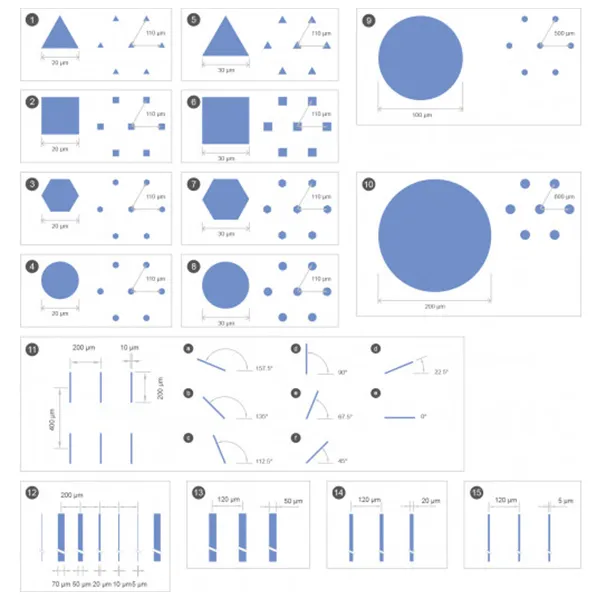
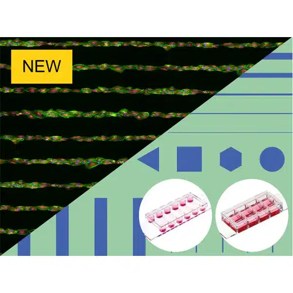
พื้นผิวไมโครแพทเทิร์นที่มีหลายรูปทรงสำหรับการทดสอบรูปแบบต่างๆ สำหรับการสร้างการทดสอบการเพาะเซลล์
- 15 รูปแบบที่แตกต่างกันบนพื้นผิวที่ไม่ยึดติดแบบพาสซีฟเพื่อการเริ่มต้นที่ง่ายดายในด้านของ micropatterning
- จัดการได้ง่ายโดยไม่ต้องเตรียม: แกะกล่องและเริ่ม
- ห้องสร้างภาพคุณภาพเชิงแสงที่ยอดเยี่ยมสำหรับกล้องจุลทรรศน์ความละเอียดสูง
การใช้งาน
- การกำหนดรูปทรงไมโครแพทเทิร์นที่เหมาะสมสำหรับการประยุกต์ใช้การเพาะเลี้ยงเซลล์ที่คุณเลือก
- การเพาะเลี้ยงและการสร้างภาพเซลล์มวลรวมสามมิติหรือโมโนเลเยอร์ของแพทช์เซลล์
- การเพาะเลี้ยงและการวิเคราะห์ด้วยกล้องจุลทรรศน์ของทรงกลม, ออร์แกนอยด์, ตัวอ่อนและเซลล์ต้นกำเนิด
- การวิเคราะห์เซลล์เดี่ยวโดยใช้วิธีการต่างๆ (เช่น การทรานส์เฟกชัน โปรตีโอมิกส์ หรือการทดสอบกิจกรรมการเผาผลาญ) ด้วยการอ่านค่าด้วยกล้องจุลทรรศน์
- การสอบวิเคราะห์ความแปรปรวนของเซลล์เดียว (เช่น การสอบวิเคราะห์การออกฤทธิ์ของเซลล์ CAR-T)
- การเพาะเลี้ยงการรวมตัวของเซลล์ 3 มิติภายใต้การแพร่กระจายและความเค้นเฉือน (โดยใช้รูปแบบผลิตภัณฑ์ µ-Slide VI 0.4)
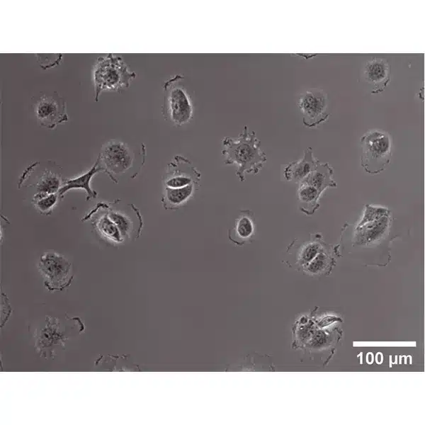
- การถ่ายภาพเซลล์แบบสดและกล้องจุลทรรศน์เรืองแสง
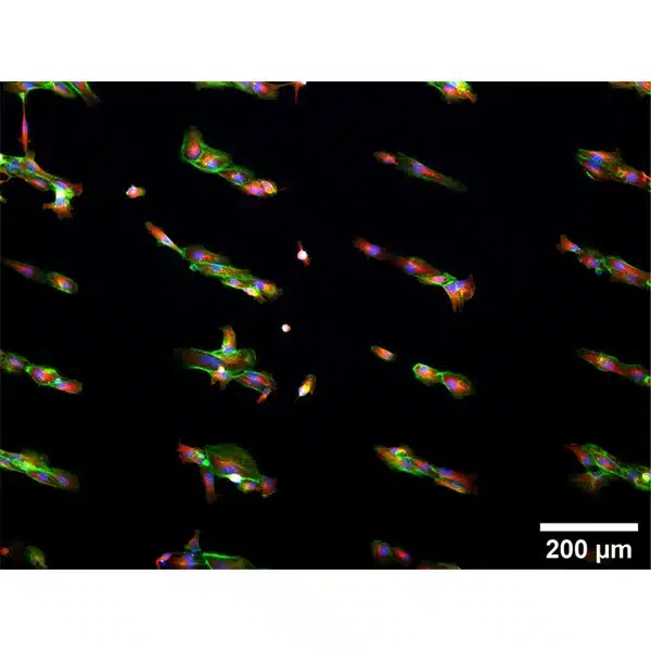
- การย้อมสีอิมมูโนฟลูออเรสเซนส์และกล้องจุลทรรศน์เรืองแสงที่มีความละเอียดสูงของเซลล์ที่มีชีวิตและเซลล์ตายตัว
A micropatterned surface with multiple geometries for testing different patterns for the establishment of cell culture assays
- 15 different patterns on a passivated, non-adhesive surface for an easy start into the field of micropatterning
- Easy handling without preparation: Unpack and start
- Excellent optical-quality imaging chamber for high-resolution microscopy
Applications
- Determination of a suitable micropatterning geometry for your chosen cell culture application
- Culture and imaging of three-dimensional cell aggregates or cell patch monolayers
- Culture and microscopic analysis of spheroids, organoids, embryoid bodies, and stem cells
- Analysis of single cells using various approaches (e.g., transfection, proteomics, or metabolic activity tests) with microscopy readout
- Single-cell variability assays (e.g., CAR-T cell activity assay)
- Culture of 3D cell aggregates under perfusion and shear stress (using the µ-Slide VI 0.4 product variation)
- Live cell imaging and fluorescence microscopy
- Immunofluorescence staining and high-resolution fluorescence microscopy of living and fixed cells
