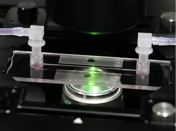
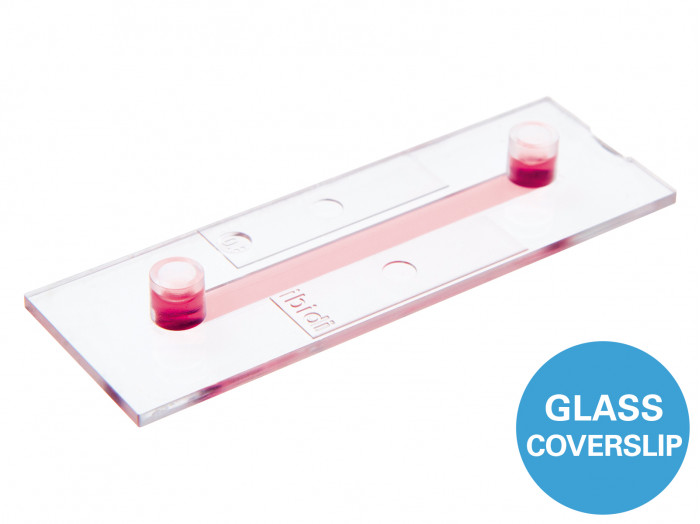
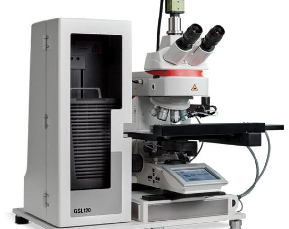
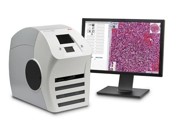
Channel slides with a #1.5H glass bottom for flow applications; different heights and volumes available
- Large area of uniform shear stress
- Easy connection using Luer adapters
- Homogeneous cell distribution with defined optical pathway
- Super-resolution and TIRF possible due to the #1.5H glass substrate
- Surface Modification: #1.5H (170 µm +/- 5 µm) D 263 M Schott glass, sterilized
Pcs./Box: 15 (individually packed)
Channel Version (Channel Height): 0.2 (Channel Height 250 µm) - Surface Modification: #1.5H (170 µm +/- 5 µm) D 263 M Schott glass, sterilized
Pcs./Box: 15 (individually packed)
Channel Version (Channel Height): 0.4 (Channel Height 450 µm) - Surface Modification: #1.5H (170 µm +/- 5 µm) D 263 M Schott glass, sterilized
Pcs./Box: 15 (individually packed)
Channel Version (Channel Height): 0.6 (Channel Height 650 µm) - Surface Modification: #1.5H (170 µm +/- 5 µm) D 263 M Schott glass, sterilized
Pcs./Box: 15 (individually packed)
Channel Version (Channel Height): 0.8 (Channel Height 850 µm)
Applications
- Adherent cells under flow conditions
- Cell culture (static or stop-flow)
- Simulation of blood vessels with endothelial cells
- High-resolution microscopy of living and fixed cells
- TIRF and super-resolution microscopy
Want to know if you should use a glass or a polymer bottom for your application? Find out here.
Specifications
| Outer dimensions | 25.5 x 75.5 mm² (w x l) |
| Channel length | 50 mm |
| Channel width | 5 mm |
| Adapters | Female Luer |
| Volume per reservoir | 60 μl |
| Growth area | 2.5 cm2 |
| Bottom: Glass coverslip No. 1.5H, selected quality, 170 µm +/- 5 µm | |

Because of the adhesive layer, the channel height of the µ-Slide I Luer Glass Bottom increases by 50 µm compared to the µ-Slide I Luer.
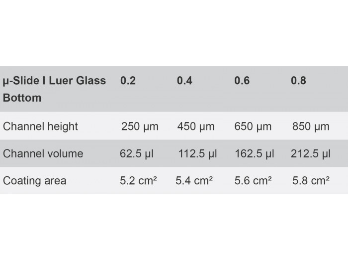
| µ-Slide I Luer Glass Bottom | 0.2 | 0.4 | 0.6 | 0.8 |
| Channel height | 250 µm | 450 µm | 650 µm | 850 µm |
| Channel volume | 62.5 µl | 112.5 µl | 162.5 µl | 212.5 µl |
| Coating area | 5.2 cm² | 5.4 cm² | 5.6 cm² | 5.8 cm² |

Find Out More
Please find more detailed information about the planning, conduction, and data analysis of cell culture under flow assays here.
Download the whole “Cell Culture Under Flow” Application Guide as a PDF here.
Technical Features
- Standard format with thin coverslip bottom made from D 263 M Schott glass #1.5H (170 µm +/- 5 µm) for low or high magnification microscopy
- Large observation area for microscopy
- Defined shear stress and shear rate levels
- Easy connection to tubes and pumps using Luer adapters
- Fully compatible with the ibidi Pump System
- May require coating to promote cell attachment
- Also available as a µ-Slide I Luer with an ibidi Polymer Coverslip Bottom for superior cell growth

The Principle of the µ-Slide I Luer Glass Bottom
The Glass Bottom
The µ-Slide I Luer Glass Bottom comes with a thin #1.5H Glass Coverslip Bottom that has the highest optical quality and is ideally suitable for high-resolution microscopy, TIRF, and super-resolution microscopy. To promote cell attachment, a surface coating might be required prior to cell seeding.
Find more information and technical details about the coverslip bottom of the ibidi chambers here.

Choosing the Right Channel Height for Flow Applications
Which Experiment Are You Planning?
Low channels are more suitable for flow applications.
High channels are more suitable for static cell culture.
| For flow assays with small amounts of medium and high values of shear stress | 0.2 channel |
| For a wide range of shear stress | 0.4 channel |
| For controlling low values of shear stress < 2 dyne/cm2 | 0.6 and 0.8 channels |
Select the ideal Perfusion Set for your flow application here.
Cross Section of the Different Channel Heights

* not recommended for static culture over more than 6 hours
Experimental Example
Cells Cultured Under Static or Flow Conditions
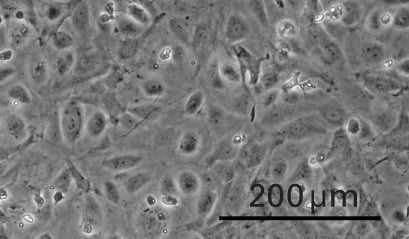
Static cultivation
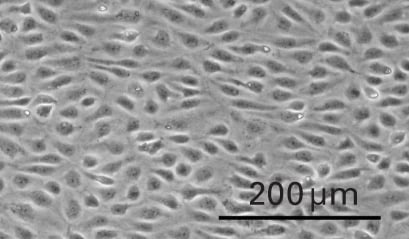
Flow cultivation
