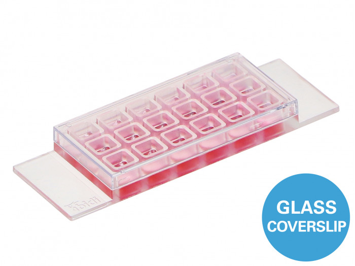
ibidi : µ-Slide 18 Well Glass Bottom
ฝาครอบกระจกแชมเบอร์ 18 หลุมสำหรับการเพาะเลี้ยงเซลล์ เหมาะสำหรับกับกล้องจุลทรรศน์ TIRF และการใช้งานโมเลกุลเดี่ยว
- สไลด์ 18 หลุมแบบ All-in-one สำหรับการทดลองที่คุ้มต้นทุนด้วยตัวเลขเซลล์ขนาดเล็กและปริมาณรีเอเจนต์ต่ำ
- สไลด์ไมโครสโคปที่เหมาะสำหรับการถ่ายภาพความละเอียดสูงผ่านก้นแก้ว No. 1.5H ที่มีคุณภาพด้านออปติคอลสูงสุดและความหนาผันแปรต่ำ
- ห้องเพาะเลี้ยงเซลล์เหมาะสำหรับเทคนิคกล้องจุลทรรศน์ส่วนใหญ่ (เช่น TIRF, กล้องจุลทรรศน์ความละเอียดสูง, DIC, การเรืองแสงแบบไวด์ฟิลด์, กล้องจุลทรรศน์แบบคอนโฟคอล, กล้องจุลทรรศน์แบบสองโฟตอน, FRAP, FRET, FLIM และ LSFM)
การใช้งาน
- การปลูกถ่ายและกล้องจุลทรรศน์ความละเอียดสูงของเซลล์
- TIRF และการประยุกต์ใช้โมเลกุลเดี่ยวในเซลล์ที่มีชีวิตและเซลล์ตายตัว
- กล้องจุลทรรศน์ความละเอียดสูง
- การย้อมสีด้วยอิมมูโนฟลูออเรสเซนส์และกล้องจุลทรรศน์เรืองแสงของเซลล์ที่มีชีวิตและเซลล์ตายตัว
- การสร้างภาพเซลล์แบบสดในช่วงเวลาที่ขยายออกไป
- การตรวจการถ่ายเท
- กล้องจุลทรรศน์แบบดิฟเฟอเรนเชียลรบกวนคอนทราสต์ (DIC) เมื่อใช้กับฝาปิด DIC
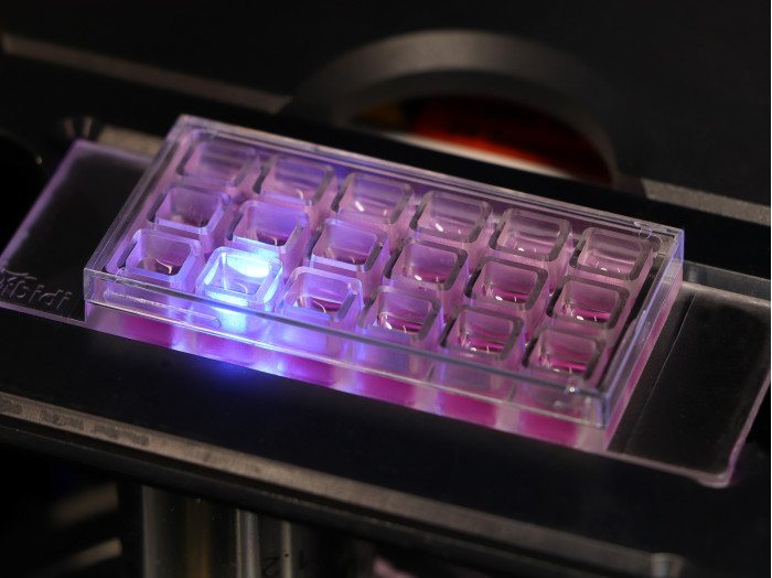
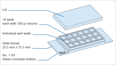
A chambered glass coverslip with 18 wells for cell culture, suitable for use in TIRF microscopy and single molecule applications
- All-in-one 18 well chamber slide for cost-effective experiments with small cell numbers and low reagent volumes
- Microscopy slide that is ideal for high-resolution imaging through the No. 1.5H glass bottom with the highest optical quality and low thickness variability
- Cell culture chamber suitable for most microscopy techniques (e.g., TIRF, super-resolution microscopy, DIC, widefield fluorescence, confocal microscopy, two-photon microscopy, FRAP, FRET, FLIM, and LSFM)
- Surface Modification: #1.5H (170 µm +/- 5 µm) D 263 M Schott glass, sterilized
Pcs./Box: 15 (individually packed)
Applications
- Cultivation and high-resolution microscopy of cells
- TIRF and single molecule applications of living and fixed cells
- Super-resolution microscopy
- Immunofluorescence staining and fluorescence microscopy of living and fixed cells
- Live cell imaging over extended time periods
- Transfection assays
- Differential interference contrast (DIC) microscopy when used with a DIC lid
Specifications
| Outer dimensions (w x l) | 25.5 x 75.5 mm² |
| Number of wells | 18 |
| Dimensions of wells (w x l x h) | 5.7 x 6.1 x 6.8 mm³ |
| Volume per well | 100 µl |
| Height with/without lid | 8.2/6.8 mm |
| Growth area per well | 0.34 cm² |
| Coating area per well | 1.15 cm² |
| Bottom: Glass coverslip No. 1.5H, selected quality, 170 µm +/- 5 µm | |
Technical Features
- Chambered glass coverslip with 18 independent wells
- Bottom made from D 263 M Schott glass, No. 1.5H (170 +/- 5 µm)
- May require coating to promote cell attachment
- Imaging chamber slide with excellent optical quality for high-end microscopy
- Closely fitting lid for low evaporation
- Individual well walls for minimizing well-to-well crosstalk and contaminations
- Compatible with staining and fixation solutions
- Also available with an ibidi Polymer Coverslip Bottom for superior cell growth: µ-Slide 18 Well
- Also available as an adhesive version without a bottom: sticky-Slide 18 Well
Application Examples
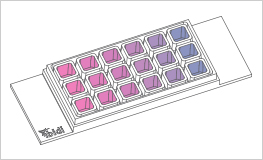
The µ-Slide 18 Well is ideal for immunofluorescence stainings, toxicological screenings, and cell surface coatings, especially, when multiple conditions on one slide are needed.
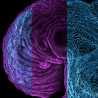
Immunofluorescent staining of human intestinal organoids in the ibidi µ-Slide 18 Well Glass Bottom. The organoids are derived from induced pluripotent stem cells and illustrate the beauty of their well-organized structures. Composition of two images showing cell nuclei (blue, DAPI) and F-actin (magenta, phalloidin). The picture was acquired using a Zeiss LSM 780 confocal microscope with a 10x objective. Image by Veronika Bosáková, Jan Frič, and Marco De Zuani, Cellular and Molecular Immunoregulation, International Clinical Research Center, St. Anne’s University Hospital, Brno, Czech Republic.
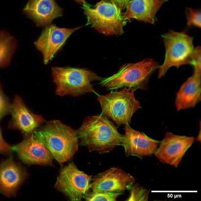
Fluorescence microscopy of rat fibroblasts (Rat1) in a µ-Slide 18 Well Glass Bottom. Red: alpha-tubulin, green: F-actin, stained with LifeAct-TagGFP2 Protein; blue: nuclei (ibidi Mounting Medium with DAPI). 60x
