ibidi : µ-Dish 35 mm, high – จานเพาะเลี้ยงเซลล์ สำหรับการถ่ายภาพ ขนาด 35 มม. (ขอบสูง)
จานเพาะเลี้ยงเซลล์ แบบก้นบางพิเศษ แบบ polymer coverslip สำหรับการถ่ายภาพ เส้นผ่านศูนย์กลางขนาด 35 มม. – ให้ภาพความละเอียดสูง เหมาะสำหรับยึดติด หรือ Adherence cell
มีเทคโนโลยี ibiTreat surface เพื่อการเจริญเติบโตของเซลล์ มีฝาปิด พร้อมตำแหน่งล็อคที่ขอบจาน ช่วยลดการระเหยเหมาะสำหรับเทคนิค DIC เมื่อใช้ร่วมกับฝาปิดพิเศษสำหรับเทคนิค DIC
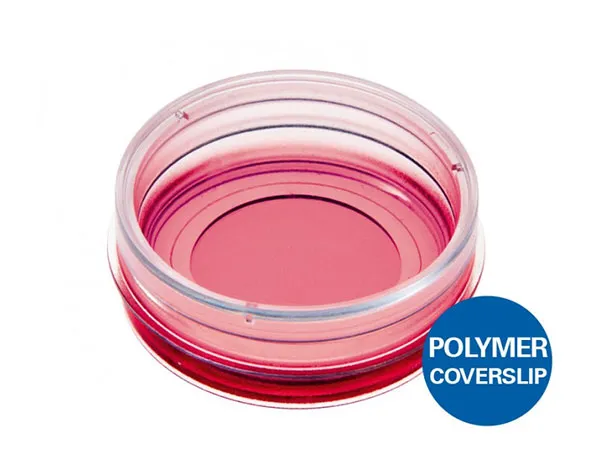
A 35 mm imaging dish with a polymer coverslip bottom for high-end microscopy and cell-based assays
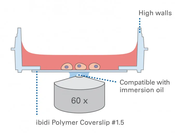
- In this microscopy dish, the cells are imaged on a No. 1.5 ibidi Polymer Coverslip bottom with the highest optical quality and optimal cell adherence
- Cell culture petri dish for everyday use with diverse application possibilities; equipped with a lid with locking feature that minimizes evaporation
- Suitable for DIC when using a DIC Lid
- Surface Modification: Uncoated: #1.5 polymer coverslip, hydrophobic, sterilized
Pcs./Box: 60 (individually packed) - Surface Modification: ibiTreat: #1.5 polymer coverslip, tissue culture treated, sterilized
Pcs./Box: 400 (individually packed)
Applications
- Cultivation and high-resolution imaging of cells
- Immunofluorescence staining
- Widefield and confocal fluorescence microscopy of living and fixed cells
- Live cell imaging over extended time periods
- Transfection assays
- Differential interference contrast (DIC) when using a DIC Lid
- Cell location and counting when using the gridded version
Want to know if you should use a glass or a polymer bottom for your application? Find out here.
Specifications
| Ø µ-Dish | 35 mm |
| Volume | 2 ml |
| Growth area | 3.5 cm2 |
| Coating area using 400 µl | 4.1 cm2 |
| Ø observation area | 21 mm |
| Height with / without lid | 14/12 mm |
| Bottom: ibidi Polymer Coverslip | |
Technical Features
- Standard format imaging dish with a 35 mm diameter for tissue culture
- ibiTreat (tissue culture-treated) surface for optimal cell adhesion and cell growth
- High walls with a standard height for easy handling; low wall version available: µ-Dish 35 mm, low
- Lid with locking feature for minimal evaporation
- Rim for easy opening
- No autofluorescence
- Biocompatible plastic material—no glue, no leaking
- Available as a Bulk Pack with 400 individually packed µ-Dishes per box
- Available with a #1.5H Glass Coverslip Bottom: µ-Dish 35 mm, high Glass Bottom, specifically suitable for TIRF and single molecule applications
- Available with a 500 µm grid for cell counting: µ-Dish 35 mm, high Grid-500
- Available with a non-adhesive Bioinert surface: µ-Dish 35 mm, high Bioinert, specifically suitable for 3D applications, such as spheroids and suspension cells
Test our everyday solution for cost-effective high-throughput experiments: Glass Bottom Dish 35 mm

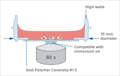
The Principle of the µ-Dish 35 mm, high
The Coverslip Bottom
The µ-Dish 35 mm, high comes with a thin ibidi Polymer Coverslip Bottom that has the highest optical quality (comparable to glass) and is ideally suitable for high-resolution microscopy. It is also available with a #1.5H Glass Coverslip Bottom for TIRF or single molecule microscopy.
As another option for cost-effective high-throughput experiments, we offer the Glass Bottom Dish 35 mm with a #1.5 glass coverslip bottom.
Find more information and technical details about the coverslip bottom of the ibidi chambers here.
Find a comparison of the different ibidi Dishes 35 mm here.
The ibiTreat Surface
ibiTreat (tissue culture-treated) is our most recommended surface modification, because almost all adherent cells grow well on it without the need for any additional coating.
Find more information about the different surfaces of the ibidi chambers here.
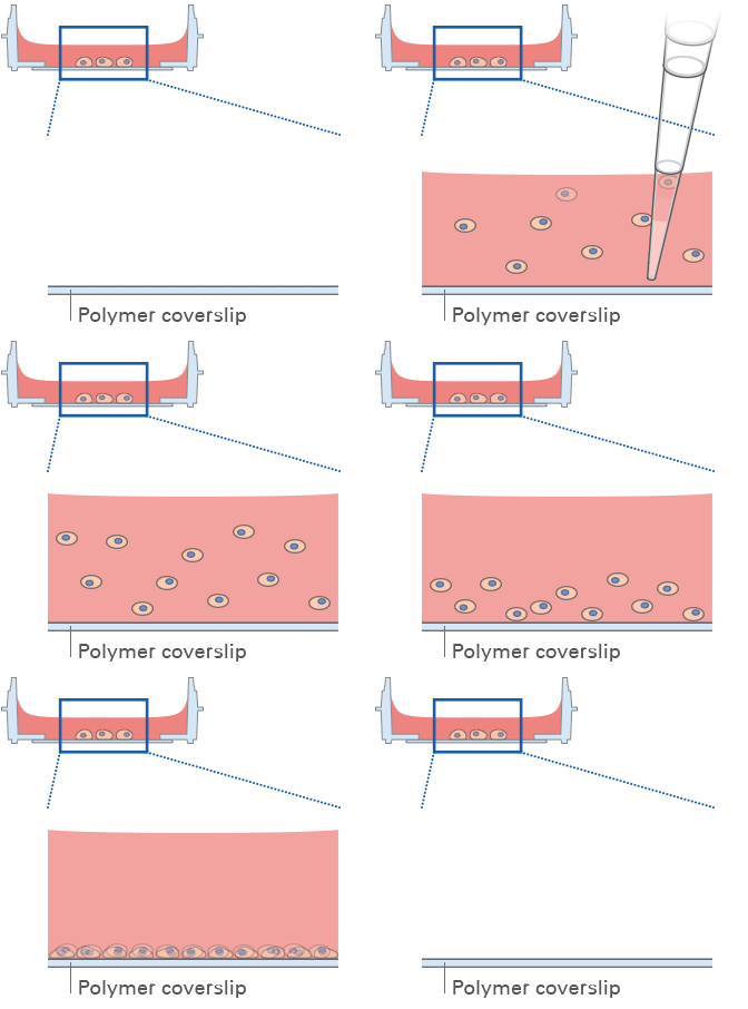
Lid with Locking Feature for Minimized Evaporation
All ibidi µ-Dishes are equipped with the special lid-locking feature. The locking position minimizes evaporation, and thereby provides excellent conditions for long-term studies in a non-humidified environment. Gas exchange (carbon dioxide or oxygen) during cell culture is maintained thanks to the gas-permeable plastic material of the dish.
TIP: Use the locking feature if minimal evaporation is required (e.g., outside incubators, non-humidified microscopy stages, etc.).

Experimental Examples
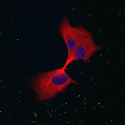
Differentiated PC12 cells in a μ-Dish 35 mm, high, stained for ß-III-Tubulin (Covance, Princeton, USA) in red. PC12 cells were incubated with commercial green-fluorescent magnetic nanoparticles (MAG-ARA, Chemicell, Berlin) and nuclei were stained with 4‘, 6-diamidino-2- phenylindole (DAPI, Roche Applied Systems, Indianapolis, USA). Image by Josephine Pinkernelle, Otto-Von-Guericke University Magdeburg, Magdeburg, Germany.
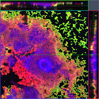
Ultra-structure of Streptococcus pyogenes biofilm grown on mouse embryonic fibroblasts in a μ-Dish 35 mm, high ibiTreat. Blue – Phalloidin AF350; Green – anti-Streptococcus pyogenes primary antibody, Rabbit anti-goat AF488 secondary antibody; Red – WGA AF594. Image by Anuradha Vajjala, Singapore Centre on Environmental Life Sciences Engineering, Nanyang Technological University, Singapore.
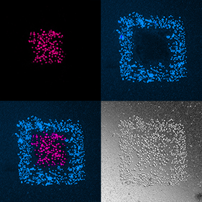
Brightfield and fluorescence images of multicellular shapes created in the μ-Dish 35 mm, high using Biopixlar, a bioprinter capable of generating multicellular biological tissues directly in the cell culture media. Images by Fluicell.
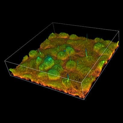
Live rat kidney cells stained with Di-8-ANEPPS to image primary cilia. Cells were plated on a μ-Dish and imaged on a Nikon A1R inverted confocal microscope with a 60X NA 1.27 WI objective using 457nm excitation and a 490nm LP filter to the detection PMT. 3D volume rendering has been depth coded with NIS Elements software. Image by Brian Siroky, Alyssa Wilson, and Matthew Kofron, Cinncinnati Children’s Hospital, Cincinnati, OH, USA.
ibidi µ-Dish Height Variations
µ-Dish 35 mm, high


High walls for all standard cell culture applications


Low walls that provide greater cell access, which is useful for micromanipulation
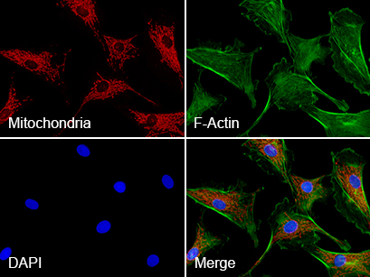
Triple immunofluorescence of bovine endothelial cells.
Red: mitochondria, stained with MitoTracker™ Red CMXRos;
Green: F-actin, stained with Alexa Fluor™ 488 Phalloidin;
Blue: nuclei, stained with DAPI.
Comparison of the ibidi Dishes 35 mm
Please find a detailed comparison of the material specifications, including suitable microscopy applications for the ibidi Polymer Coverslip, the ibidi Glass Bottom, and further materials here.
| Bottom material | #1.5 ibidi Polymer Coverslip | #1.5H ibidi Glass Coverslip | #1.5 glass coverslip |
| Bottom thickness | 180 µm (+10/-5 µm) | 170 µm (+/-5 µm) | 170 µm (+20/-10 µm) |
| Bottom: gas permeability | Yes | No | No |
| Available surfaces | ibiTreat (tissue culture treated), Uncoated (hydrophobic) | Uncoated glass | Uncoated glass |
| Lid | Lid with locking feature | Lid with locking feature | Standard lid |
| Packaging | Sterile, individually packed | Sterile, individually packed | Sterile, 10 pcs. per case |
| Quantity | 60 or 400 pcs./box | 60 or 400 pcs./box | 200 or 800 pcs./box |
| Low wall version available? | Yes: µ-Dish 35 mm, low | Yes: µ-Dish 35 mm, low Glass Bottom | No |
| Gridded version available? | Yes: µ-Dish 35 mm, high Grid-500 | Yes: µ-Dish 35 mm, high Grid-50 Glass Bottom, µ-Dish 35 mm, high Grid-500 Glass Bottom | No |
