ibidi : µ-Slides With Multi-Cell µ-Pattern
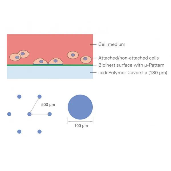
พื้นผิวไมโครแพทเทิร์นสำหรับการทดสอบหลายเซลล์ด้วยการอ่านค่าด้วยกล้องจุลทรรศน์เรืองแสง
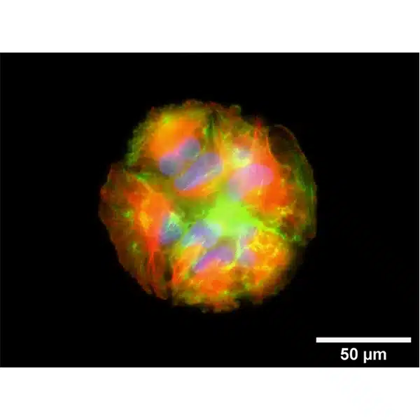
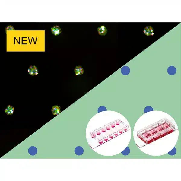
- รูปแบบหลายเซลล์ที่มีระยะห่างในอุดมคติสำหรับ 3D spheroids หรือ organoids
- ใช้งานง่ายโดยไม่ต้องเตรียม: แกะและสตาร์ท
- ห้องภาพคุณภาพเชิงแสงที่ยอดเยี่ยมสำหรับกล้องจุลทรรศน์ความละเอียดสูง
การใช้งาน
- การเพาะเลี้ยงและการสร้างภาพเซลล์มวลรวมสามมิติหรือโมโนเลเยอร์ของแพทช์เซลล์สองมิติ
- การเพาะเลี้ยงและการวิเคราะห์ทรงกลม ออร์กานอยด์ ตัวอ่อน และสเต็มเซลล์ด้วยการอ่านค่าด้วยกล้องจุลทรรศน์
- การเพาะเลี้ยงการรวมตัวของเซลล์ 3 มิติภายใต้การแพร่กระจายและความเค้นเฉือน (โดยใช้รูปแบบผลิตภัณฑ์ µ-Slide VI 0.4)
- การทดสอบสิ่งที่แนบมาของประเภทเซลล์ของคุณบนแม่ลายการผูก RGD ด้วย Trial Pack
- การถ่ายภาพเซลล์แบบสดและกล้องจุลทรรศน์เรืองแสง
- การย้อมสีอิมมูโนฟลูออเรสเซนส์และกล้องจุลทรรศน์เรืองแสงที่มีความละเอียดสูงของเซลล์ที่มีชีวิตและเซลล์ตายตัว
A micropatterned surface for multi-cell assays with fluorescence microscopy readout
- Multi-cell pattern with ideal spacing for 3D spheroids or organoids
- Easy handling without preparation: Unpack and start
- Excellent optical-quality imaging chamber for high-resolution microscopy
Applications
- Culture and imaging of three-dimensional cell aggregates or two-dimensional cell patch monolayers
- Culture and analysis of spheroids, organoids, embryoid bodies, and stem cells with microscopy readout
- Culture of 3D cell aggregates under perfusion and shear stress (using the µ-Slide VI 0.4 product variation)
- Testing the attachment of your cell type on the RGD binding motif with the Trial Pack
- Live cell imaging and fluorescence microscopy
- Immunofluorescence staining and high-resolution fluorescence microscopy of living and fixed cells

