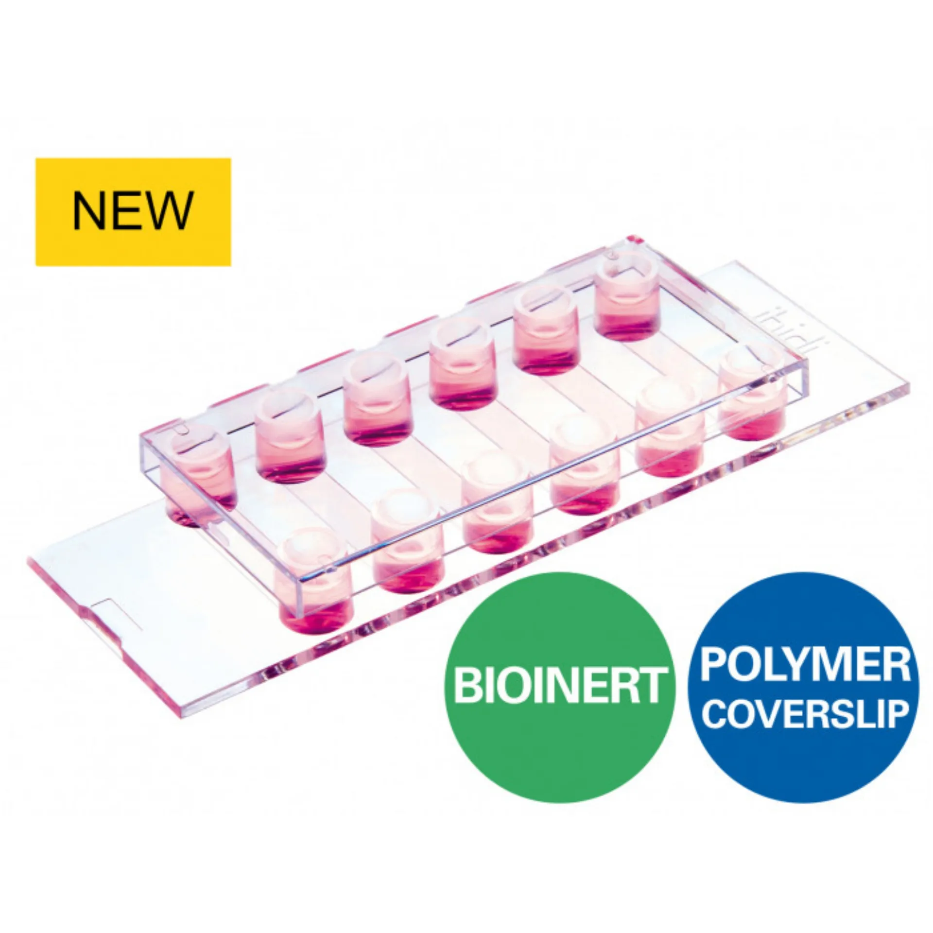
ibidi : µ-Slide VI 0.4 Bioinert
A 6 channel μ-Slide ที่มีพื้นผิวที่ไม่ยึดเกาะสำหรับการทดลองการไหลและกล้องจุลทรรศน์เรืองแสง
- เหนือกว่าพื้นผิวที่มีการยึดเกาะต่ำเป็นพิเศษ (ULA) มาตรฐาน—พื้นผิวเฉื่อยทางชีวภาพที่เสถียรสำหรับการทดลองระยะยาวโดยไม่ยึดเกาะกับเซลล์หรือชีวโมเลกุลใดๆ
- คุณภาพระดับจุลทรรศน์ที่ยอดเยี่ยมโดยไม่มีการเรืองแสงอัตโนมัติ— ใช้พื้นผิวร่วมกัน 100% มีคุณภาพแสงสูงสุดของ ibidi : Polymer Coverslip
การใช้งาน
- การถ่ายภาพแบบเรียลไทม์ภายใต้สภาวะคงที่หรือสภาวะการไหล
- การสร้างและการเพาะเลี้ยงเซลล์ 3 มิติในระยะยาว (เช่น ทรงกลมและออร์แกนอยด์) โดยไม่มีโครง
- กล้องจุลทรรศน์เรืองแสงความละเอียดสูงของออร์กานอยด์ สเฟียรอยด์ ตัวอ่อน (EBs) และเซลล์ในช่วงล่าง
- การวิเคราะห์ปฏิสัมพันธ์ระหว่างเซลล์และเซลล์โดยปราศจากเบื้องหลัง
- ป้องกันการแตกตัวของสเต็มเซลล์เนื่องจากการยึดติด
- การเพาะเลี้ยงเซลล์แขวนลอยในสถานะไม่ผูกมัดอย่างถาวร
- การทดสอบการก่อตัวของเนื้องอกทรงกลมด้วยตนเอง
- โมเดลเนื้องอกทรงกลม 3 มิติ
A 6 channel μ-Slide with a non-adherent surface for flow experiments and fluorescence microscopy
- Superior to the standard ultra-low attachment (ULA) surfaces—stable, biologically inert surface for long-term experiments without any cell or biomolecule adhesion
- Excellent microscopic quality without any autofluorescence—100% surface passivation combined with the highest optical quality of the ibidi Polymer Coverslip
Applications
- Real-time imaging under either static or flow conditions
- Generation and long-term culture of 3D cell aggregates (e.g., spheroids and organoids) without any scaffold
- High resolution fluorescence microscopy of organoids, spheroids, embryoid bodies (EBs), and cells in suspension
- Background-free analysis of cell-cell interactions
- Prevention of stem cell differentiation due to attachment
- Culture of suspension cells in a permanently unattached state
- Self-assembly tumor spheroid formation assays
- 3D tumor spheroid models
