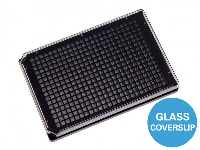
ไมโครเพลทจำนวน 384 หลุมสีดำชนิดก้นทำจากแก้ว
A black 384 well plate with square wells and a #1.5H glass coverslip bottom for high throughput screening using TIRF and super-resolution microscopy
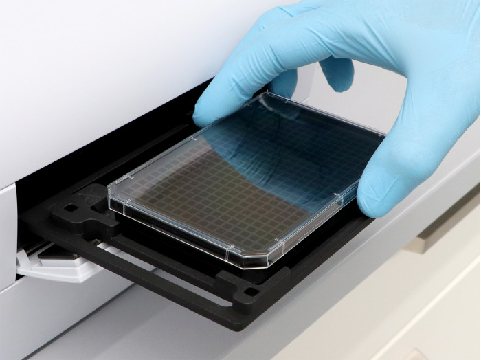
- Compatible with robotics, due to an ANSI/SLAS (SBS) standard format
- Brilliant cell imaging due to the low thickness variability of the #1.5H glass coverslip and the excellent inner well and whole plate flatness
- Low well-to-well crosstalk in fluorescence microscopy
- Surface Modification: #1.5H (170 µm +/- 5 µm) D 263 M Schott glass, sterilized
Pcs./Box: 15 (individually packed)
Applications
- Cultivation, high throughput screening (HTS), and high-resolution microscopy of cells
- Widefield fluorescence imaging and confocal microscopy of living and fixed cells
- Compound screenings (toxicology)
- Large-scale transfection experiments
- Live cell imaging, also suited for extended periods
- Total internal reflection fluorescence (TIRF) and single molecule applications
- Super-resolution microscopy (STED, SIM, (F)PALM, (d)STORM) and fluorescence correlation spectroscopy (FCS)
- Immunofluorescence staining
Specifications
| Length / width | 127.7 / 85.5 mm |
| Height with / without lid | 17.2 / 15.0 mm |
| Well-to-well distance | 4.5 mm |
| Well clearance | 0.73 mm |
| Single well dimensions | 3.4 x 3.4 mm² |
| Single well depth | 12.8 mm |
| Volume per well | 50 µl |
| Growth area per well | 0.11 cm² |
| Coating area per well using 50 µl | 0.8 cm² |
| Bottom: Glass Coverslip No. 1.5H, selected quality, 170 µm +/- 5 µm | |
Technical Drawing
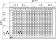 | Technical drawings and details are available in the Instructions (PDF). |
Technical Features
- Standard format, compatible with robotics and plate readers due to an ANSI/SLAS (SBS) standard format
(85.5 x 127.5 mm) - 384 quadratic wells with standard numbering
(letters A–P and numbers 1–24) - Suitable for fluorescence scanners, plate readers, robotics, and lab automation
- Bottom made from D 263 M Schott glass with a thickness of 170 µm +/- 5 µm
- May require coating to promote cell attachment
- No autofluorescence
- Excellent inner well flatness and whole plate flatness
- Compatible with staining and fixation solvents
- Non-cytotoxic and biocompatible
- Immersion oil compatible
- Sterile with single packaging
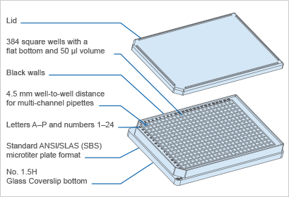
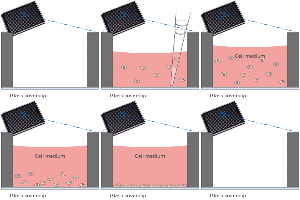
The Principle of the µ-Plate 384 Well Black Glass Bottom
The Coverslip Bottom
The µ-Plate 384 Well Black Glass Bottom features optimal flatness properties, which are crucial for high-resolution microscopy in screening applications. The glass coverslip provides high accuracy and facilitates imaging from well to well across the entire plate with less variation in focus due to the rigid material with high mechanical stability.
Application Examples

Fluorescence microscopy of immunostained Rat1 fibroblasts in the µ-Plate 384 Well Black Glass Bottom. Cells were stained for F-actin (green), alpha-Tubulin (red) and nuclei (DAPI, blue) and imaged using a 60x objective.

Composition of 16 stitched fluorescence microscopy images showing one whole well of the µ-Plate 384 Well Black Glass Bottom. Rat1 cells were stained with DAPI to visualize the nuclei and imaged using a 10x objective. Overlay with brightfield (well contours).
