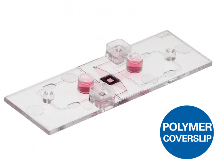
A µ-Slide with a porous silicon nitride membrane for imaging of transmigration and transport studies under flow or static conditions
- Brilliant optical quality through the thin, porous membrane
- Full access to the apical and basal sides of adherent cells in numerous applications
- Different pore sizes available for various cell types
Applications
- Trans-endothelial migration under flow conditions
- Co-cultivation of cell layers and transport assays in 2D or in a 3D gel matrix
- Apical-basal cell polarity assays
- Cell barrier model assays with apical-basal gradients
- Cell migration assays (e.g., to investigate tumor invasion or metastasis)
Selected publications:
Salvermoser, Melanie, et al. “Myosin 1f is specifically required for neutrophil migration in 3D environments during acute inflammation.” Blood, The Journal of the American Society of Hematology 131.17 (2018): 1887-1898. 10.1182/blood-2017-10-811851
Read article
Rohwedder, Ina, et al. “Src family kinase-mediated vesicle trafficking is critical for neutrophil basement membrane penetration.” Haematologica (2019). 10.3324/haematol.2019.225722
Read article

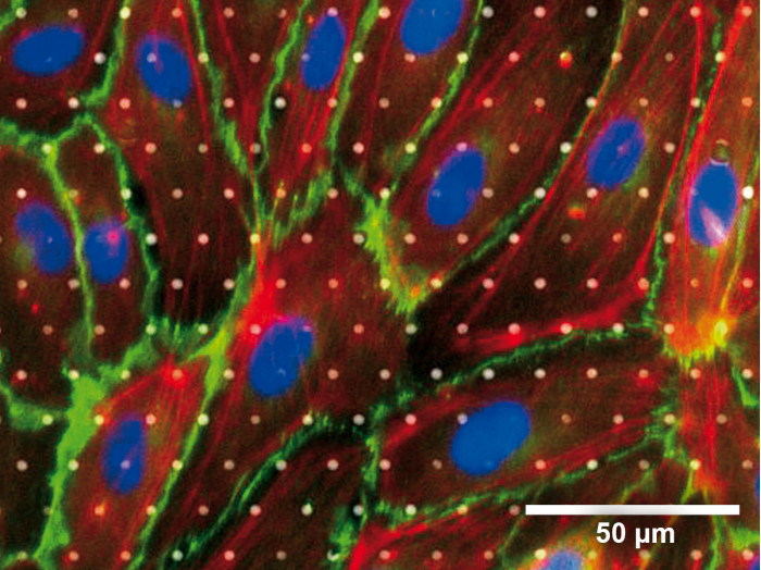
Immunofluorescence staining of human endothelial cells in the µ-Slide ibiPore, 3 µm pore size, with collagen IV coating. Overlay image of phase contrast, DAPI (blue), VE-cadherin (green), and F actin (red).
Specifications
| Total coating area | 4.50 cm² |
| Bottom | ibidi Polymer Coverslip |
| Main Channel (Lower Channel) | |
| Access | Luer port, accessible with female Luers |
| Volume | 50 µl |
| Height | 0.4 mm |
| Length | 25 mm |
| Width | 5 mm |
| Growth area | 1.25 cm² |
| Upper Channel | |
| Access | Reservoir port, accessible with 20/200 µl pipet tips |
| Volume | 55 µl |
| Height over membrane | 1.3 mm |
| ibiPore Membrane | |
| Material | Silicon nitride (SiN) |
| Thickness | 0.4 µm (400 nm) |
| Membrane size | 2 mm x 2 mm |
| Porous area | 1.77 mm x 1.84 mm |
| Restrictions for objective lenses | Working distance >0.5 mm |
| Pore layout | Hexagonal spacing |
| Available VariationsPore size 0.5 µm 3 µm 5 µm 8 µmPorosity 20% 5% 5% 5%Pore-to-pore distance 1 µm 12 µm 20 µm 32 µmWhich variation is suitable for my cell type? | |
Technical Features
- Microporous silicon nitride membranes from SiMPore Inc.
- Cross-channel structure with a porous optical membrane in between
- Excellent optical properties comparable to a glass coverslip
- Pore sizes 0.5 µm, 3 µm, 5 µm, or 8 µm available
- Membrane thickness 0.4 µm (400 nm)
- For use with objective lenses with a working distance >0.5 mm
- Fully compatible with the ibidi Pump System
- Defined shear stress and shear rate levels in the lower channel (for details see Application Note 11)

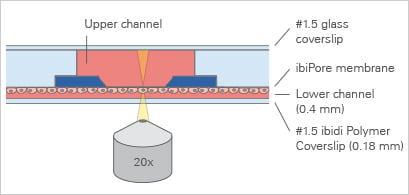
The Principle of the µ-Slide ibiPore SiN
The μ-Slide ibiPore SiN consists of a horizontal porous membrane that is inserted between two channels. The upper channel is a static reservoir above the membrane. The lower channel is a perfusion channel for applying defined shear stress on cells, which are attached to the membrane. The upper and the lower channel communicate with each other only across the membrane.
The porous membrane is made of silicon nitride (SiN), a material with very high chemical and mechanical robustness. The 400 nm thick silicon nitride membrane is ideal for imaging and microscopy purposes, without any autofluorescence or transparency issues (like glass). The SiN material can be used directly for adherent cell culture or, optionally, it can be coated with ECM proteins.

Recommended Pore Sizes and Porosities for Different Applications
What Is the Porosity?
Porosity refers to the void volume fraction of the membrane. It is defined as the volume of the pores divided by the total volume of the membrane. The graphical representations below are at the same magnification.

| Applications | Example Cell Types | Recommended Pore Sizes |
| Permeability and transport studies | Endothelial, epithelial | 0.5 µm | 3 µm |
| Cell polarity assays | Endothelial, epithelial, kidney | 0.5 µm | 3 µm |
| Co-culture assays | Fibroblasts, endothelial, epithelial, cancer, stem cells | 0.5 µm | 3 µm |
| Transmigration, chemotaxis assays | Endothelial, leukocytes (neutrophils), lymphocytes (T cells, B cells) | 3 µm | 5 µm | 8 µm |
| Invasion, migration, and metastatic potential assays | Monocytes, macrophages, lymphocytes (T cells, B cells), cancer, endothelial, epithelial, fibroblasts, osteoblasts | 3 µm | 5 µm | 8 µm |
Intended Use
These applications have been tested by the ibidi R&D team or by our customers.
Endothelial Barrier Assays
A cell monolayer is cultivated on one side of the membrane.

Cells on the lower side of the membrane

Cells on the lower side of the membrane with defined shear stress

Cells on the upper side of the membrane
Co-Culture and Cell Barrier Assay
Two separate cell monolayers are cultivated on each side of the membrane. With this method, signaling, co-culture, and transport studies are possible (e.g., analysis of drug transport across epithelial or endothelial barriers).

Co-culture of two different cell types on both sides of the membrane
Apical-Basal Cell Polarity Assays
Chemical factors inside of a 3D gel matrix lead to the polarization of a cell monolayer that is cultured on the other side of the membrane.

Cells on the lower side of the membrane with a 3D gel matrix in the upper channel

Cells on the lower side of the membrane with a 3D gel matrix with embedded cells in the upper channel
Potential Use
The following examples illustrate further potential product uses. ibidi still needs to test these applications in-house, therefore we cannot provide specific protocols. However, these applications should be possible from a technical point of view.
We would appreciate your feedback for any application of the µ-Slide ibiPore SiN that works for you. Please send your images or videos with a short description to info@ibidi.de and we will be happy to publish them on our website to support the scientific community (e.g., as User Protocol).
Trans-Membrane Migration in 2D
A cell monolayer is cultivated on one side of the membrane to observe cellular trans-membrane migration.

Transmigration of suspended leukocytes

Rolling, adhesion, and transmigration of leukocytes, for example, under defined shear stress through a monolayer of cells on the lower side of the membrane
Cell Transport in a 3D Gel Matrix
Under flow conditions, the rolling, adhesion, and transmigration of leukocytes towards chemoattractant-producing cancer cells in a 3D matrix can be observed.

Transmigration of cells across a cell monolayer on the membrane into a 3D gel matrix with embedded cells
Non-Recommended Applications
Due to technical reasons, the following applications will not work with this product and should be avoided.
This product is not intended for:
- Perfusion of the upper channel
- Perfusion of both channels
- Trans-membrane flow
- Filter applications
Application Examples
Phase Contrast Microscopy of MDCK and NIH-3T3 Cells


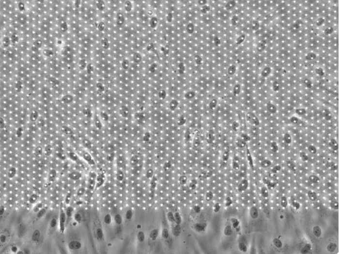
Madin-Darby canine kidney (MDCK, left) and NIH-3T3 (right) cells in the µ-Slide ibiPore SiN, 0.5 µm pore size, without any protein coating. After seeding, the cells were kept for 20 h in the incubator under static conditions. Phase contrast microscopy, 4x objective. Note that the centered, porous area in this image appears darker because the 0.5 µm pores cannot be resolved with a low-resolution objective lens.
Phase Contrast Microscopy of HUVECS Under Flow

Human umbilical vein epithelial cells (HUVEC) in the µ-Slide ibiPore SiN, 3 µm pore size, with fibronectin coating. Cells were seeded and kept for 12 h in the incubator under flow conditions (10 dyne/cm²) with the ibidi Pump System. Phase contrast microscopy after fixation, 10x objective.
Fluorescence Microscopy of the F Actin Cytoskeleton of HUVECs Under Flow

Immunofluorescence staining of human umbilical vein epithelial cells (HUVEC) in the µ-Slide ibiPore SiN, 5 µm pore size, with fibronectin coating. Cells were seeded and kept for 12 h in the incubator under flow conditions (10 dyne/cm²) with the ibidi Pump System. Green: F actin (phalloidin), blue: nuclei (DAPI). Fluorescence microscopy, 20x objective.
Decison Guide: ibidi Solutions for Two-Compartment and Membrane Assays
| µ-Slide I Luer 3D | µ-Slide ibiPore SiN | |
| Slide principle | 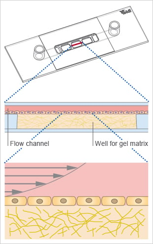 |  |
| Principle of interface | Gel-based | Membrane-based |
| Substrate for cells | Soft hydrogels (e.g., collagen or Matrigel) | Hard membrane with different pore sizes |
| Microscopy | Restricted by gel’s optics | No restriction |
| Flow application | Defined shear stress in the channel | Defined shear stress in the lower channel |
| Applications | ||
| Barrier function | Yes | Yes |
| Transmigration | Yes | Yes |
| Cell polarization | Yes | Yes |
| Shear stress | Yes | Yes |
