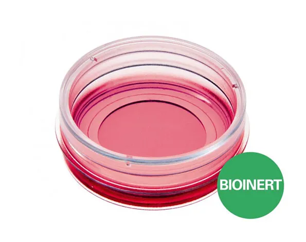
ibidi : µ-Dish 35 mm, high Bioinert – จานเพาะเลี้ยงเซลล์ชนิด Bionert ขนาด 35 มม. ขอบสูง
จานเพาะเลี้ยงเซลล์ขนาด 35 มม. พื้นผิวภาชนะป้องกันการเกาะติด – เหมาะสำหรับตัวอย่างสเฟียรอยด์, ออร์แกนอยด์ และเซลล์ลอย
- พื้นผิวชนิด ultra-low attachment (ULA) : พื้นผิวมีความเสถียร ป้องกันการยึดเยาะของเซลล์ หรือโมเลกุลใดๆ
- ไม่มีออโตฟลูออเรสเซนต์ ก้นภาชนะบางเท่า coverslip เพื่อการถ่ายภาพความละเอียดสูง
Long-term culture and high-resolution microscopy of spheroids, organoids, and suspension cells on a non-adherent surface
- Superior to the standard ultra-low attachment (ULA) surfaces: stable, biologically inert surface for long-term experiments without any cell or biomolecule adhesion
- Excellent microscopic quality without any autofluorescence: 100% surface passivation combined with the highest optical quality of the ibidi Polymer Coverslip
- Pcs./Box: 30 (individually packed)
Surface Modification: Bioinert: #1.5 polymer coverslip, surface passivation with Bioinert, sterilized
Applications
- Generation and long-term culture of spheroids and organoids without scaffold
- High resolution fluorescence microscopy of organoids, spheroids, embryoid bodies (EBs), and cells in suspension
- Background-free analysis of cell-cell interactions
- Prevention of stem cell differentiation due to attachment
- Culture of suspension cells in a permanently unattached state
- Self-assembly tumor spheroid formation assays
- 3D tumor spheroid models
Technical Features
- µ-Dish 35 mm, high with non-adherent, passivated Bioinert surface
- Ready-to-use without prior surface treatment or preparation
- 200 nm hydrophilic polyol hydrogel—stable and covalently bound
- No adsorption, coating, or binding of proteins, antibodies, enzymes, and other biomolecules
- Compatible with all fixatives and staining solutions
- No autofluorescence
- Non-cytotoxic, biologically inert, and non-degradable
- Compatible with immersion oil
Specifications
Bioinert Surface
| Bioinert Surface thickness | 200 nm |
| Bioinert Surface material | Polyol-based hydrogel |
| Protein coatings | Not possible |
µ-Dish 35 mm, high
| Ø µ-Dish | 35 mm |
| Volume | 2 ml |
| Growth area | 3.5 cm2 |
| Ø observation area | 21 mm |
| Height with / without lid | 14/12 mm |
| Bottom: ibidi Polymer Coverslip with Bioinert surface | |
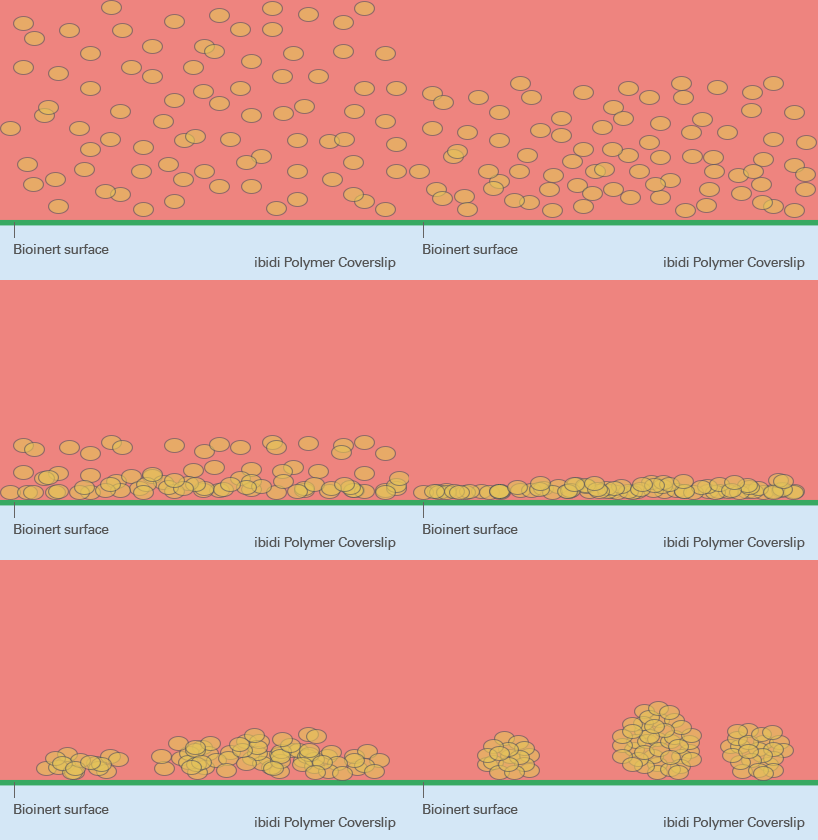
ibiTreat, Uncoated, and Bioinert—A Surface Comparison
ibiTreat
Excellent cell adhesion
- Culture of adherent cells
- ECM coatings possible
The hydrophilic ibiTreat surface provides excellent cell adhesion, even without a defined protein coating. However, ECM protein coatings can be done on ibiTreat without any restrictions. The ibiTreat surface is ideal for culture of adherent cells that do not need any specific coating.
Uncoated
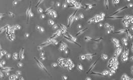
Weak cell adhesion
- Culture of adherent cells
- ECM coatings possible
The hydrophobic Uncoated surface provides weak cell adhesion, if not previously coated with an ECM protein. ECM protein coatings can be done on the Uncoated surface without any restrictions. The Uncoated surface is ideal for the culture of adherent cells that require a specific coating.
Bioinert
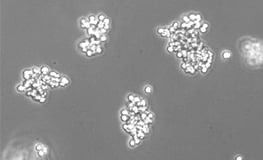
No cell adhesion
- Culture of suspension cells, cell aggregates, spheroids, embryoid bodies (EBs), organoids
- ECM coatings not possible
The hydrophilic Bioinert surface hinders any protein attachment, thus inhibiting subsequent cell attachment. Unlike with the ibiTreat and Uncoated surfaces, a coating is not possible. The Bioinert surface is ideal for the culture of suspension cells and cell aggregates.
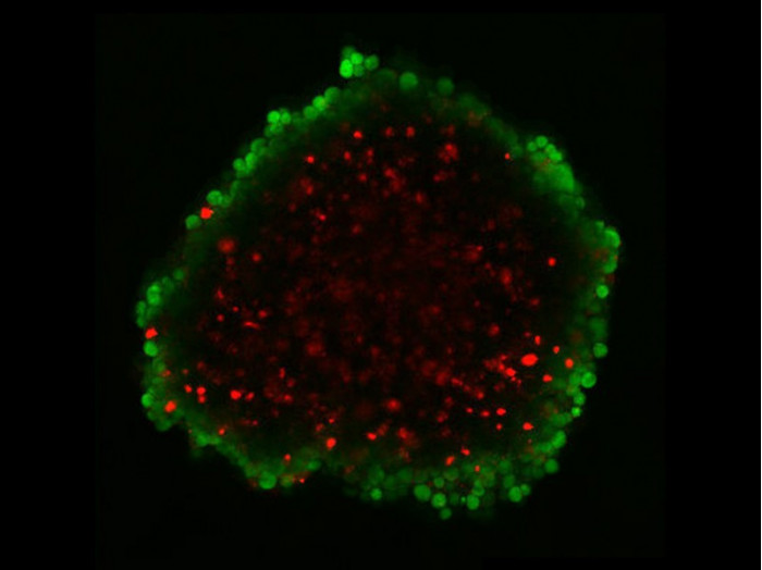
Application Example: Spheroid Culture and FDA/PI Staining Using the Bioinert Surface
The Bioinert surface is ideally suited for the short- and long-term 3D culture of spheroids and organoids. Since both specific and non-specific cell immobilization are inhibited, cells are forced into a suspended state, enabling formation of small and large spheroids. To assess the viability, the cells were stained using fluorescein diacetate (FDA), which marks viable cells, and propidium iodide (PI), which stains dead cells.
For more detailed information about FDA/PI staining, please refer to our Application Note 33, Live/Dead Staining With FDA and PI (PDF).

Confocal laser scanning section of a HepG2 spheroid. Spheroids of HepG2 cells were generated with an agarose-based liquid overlay technique and transferred to the µ-Dish 35 mm, high Bioinert. FDA (green) indicates living cells at the outer spheroid layers. PI (red) marks dead cells within the necrotic spheroid center. Microscope: Zeiss LSM 710, Plan Apochromat objective lens 10x/0.45. Scale bar is 200 µm.
