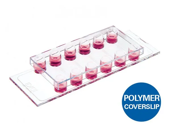
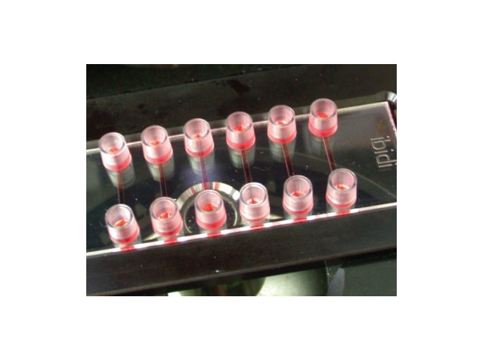
µ-Slide แบบ 6 ช่องสำหรับทำงาน flow assays แบบพร้อมกัน และสำหรับ immonofluorescence โดยมีการใช้อาหารเลี้ยงเซลล์ และ reagent ในปริมาณน้อย
- สำหรับ rolling หรือ adhesion assays ของเซลล์แบบ monolayers หรือ protein coatings
- ใช้จำนวนเซลล์ และ/หรือ ของเหลวปริมาณน้อย
- มี shear stress กลางถึงสูง
- มี 15 ชิ้น/ 1 กล่อง
A six channel µ-Slide for parallel flow assays and immunofluorescence that uses a minimal amount of reagents and medium
- Perfect for rolling and adhesion assays on cell monolayers or protein coatings
- Minimal amounts of cells and/or liquids needed
- Medium-to-high shear stress that is applicable with small volumes
- Surface Modification: Uncoated: #1.5 polymer coverslip, hydrophobic, sterilized
Pcs./Box: 15 (individually packed) - Surface Modification: ibiTreat: #1.5 polymer coverslip, tissue culture treated, sterilized
Pcs./Box: 15 (individually packed)
Applications
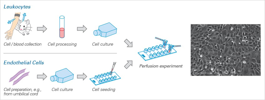
- Flow assays that use a minimum of cells and/or liquids (mouse model)
- Parallel shear stress applications
- Immunofluorescence microscopy after perfusion
- High-resolution microscopy of cells under shear stress
NOTE: Not for use in static cultures
Specifications
| Outer dimensions (w x l) | 75.5 x 25.5 mm² |
| Adapters | Female Luer |
| Number of channels | 6 |
| Channel volume | 1.7 µl |
| Channel height | 0.1 mm |
| Channel length | 17 mm |
| Channel width | 1 mm |
| Volume per reservoir | 60 µl |
| Growth area | 0.17 cm2 per channel |
| Coating area using 1.7 µl | 0.34 cm² per channel |
| Bottom: ibidi Polymer Coverslip | |
Technical Features
- Six parallel channels on one slide
- Small sample amounts
- A low dead volume
- Smallest ibidi channel slide with highest shear stress
- Easy connection to tubes and pumps using a female Luer adapter
- Defined shear stress and shear rate levels

Experimental Example
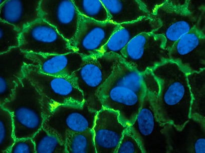
HUVEC, cultivated over 7 days at 10 dyn / cm². VE-cadherins are stained in green, cell nuclei are stained in blue.
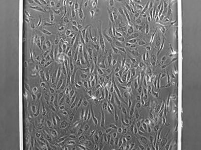
HUVEC in the µ-Slide VI 0.1 channel, cultivated over 1 day at 10 dyn / cm².
Adhesion Assay
