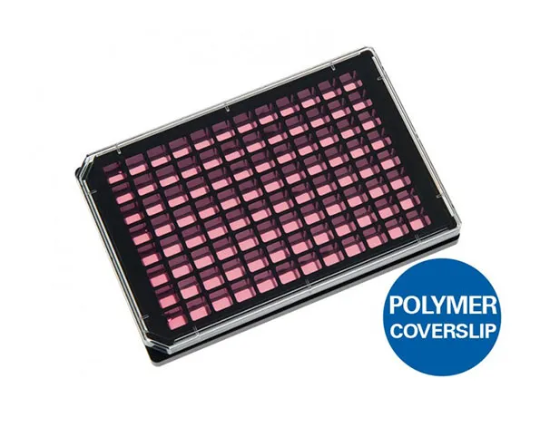
ไมโครเวลเพลทแบบก้นแบนและใสชนิด 96 หลุมแบบสี่เหลี่ยม สีดำ – สำหรับงานเพาะเลี้ยงเซลล์
- เหมาะสำหรับกล้องจุลทรรศน์
- ด้านในมีพื้นเรียบ
- ก่อให้เกิด crosstalk ต่ำในแต่ละ well ในการใช้กล้องจุลทรรศน์แบบฟลูออเรสเซนต์
A black 96 well culture plate with square wells and flat and clear bottom for high throughput microscopy
- 96 well microplate ideal for high resolution imaging through the No. 1.5 polymer coverslip bottom with the highest optical quality
- Flat bottom plate with excellent inner well flatness and whole plate flatness
- Low well-to-well crosstalk in fluorescence microscopy
- Surface Modification: Uncoated: #1.5 polymer coverslip, hydrophobic, sterilized
Pcs./Box: 15 (individually packed) - Surface Modification: ibiTreat: #1.5 polymer coverslip, tissue culture treated, sterilized
Pcs./Box: 15 (individually packed)
Applications:
- Cultivation, high throughput screening (HTS), and high-resolution microscopy of cells
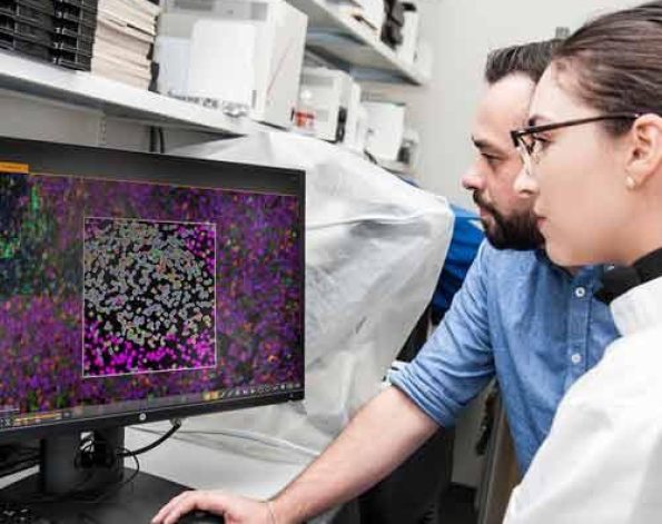
- Widefield fluorescence imaging and confocal microscopy of living and fixed cells
- Compound screenings (toxicology)
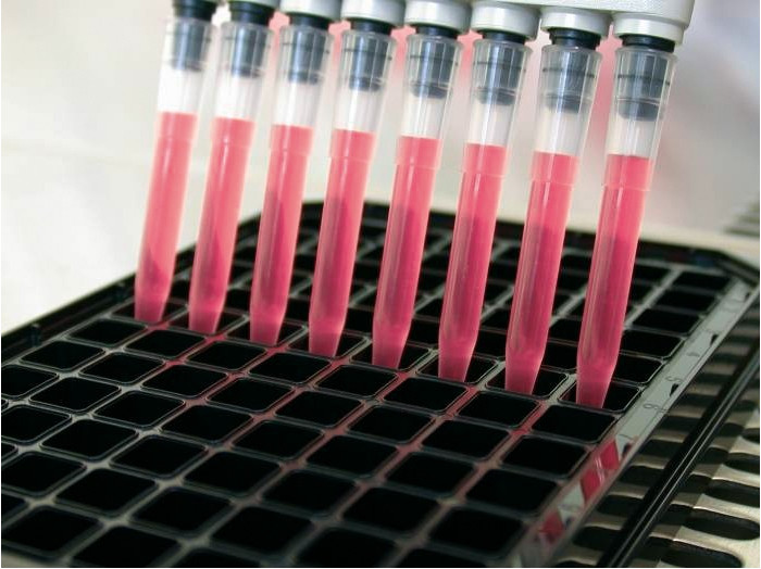
- Large-scale transfection experiments
- Live cell imaging, also suited for extended periods
- Immunofluorescence staining
Want to know if you should use a glass or a polymer bottom for your application? Find out here.
Specifications:
| Length / width | 127.8 / 85.5 mm |
| Height with / without lid | 17.2 / 15.0 mm |
| Well-to-well distance | 9.0 mm |
| Well clearance | 0.8 mm |
| Single well dimensions | 7.4 x 7.4 mm² |
| Single well depth | 12.9 mm |
| Volume per well | 300 µl |
| Growth area per well | 0.56 cm2 |
| Coating area per well using 300 µl | 2.35 cm2 |
| Bottom: ibidi Polymer Coverslip | |
Technical Drawing
 | Technical drawings and details are available in the Instructions (PDF). |
Technical Features:
- Compatible with robotics and plate readers due to an ANSI/SLAS (SBS) standard format (85.5 x 127.5 mm)
- 96 quadratic wells with standard numbering (letters A–H and numbers 1–12)
- Suitable for fluorescence scanners
- High-quality ibidi Polymer Coverslip bottom, which is compatible with solvents for staining and fixation, and also with immersion oil
- No autofluorescence
- Excellent inner well flatness and whole plate flatness
- Compatible with staining and fixation solvents
- Non-cytotoxic and biocompatible
- Compatible with immersion oil
- Sterile with single packaging
- Also available with a #1.5H Glass Coverslip Bottom: µ-Plate 96 Well Black Glass Bottom for special microscopic applications
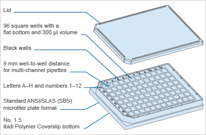
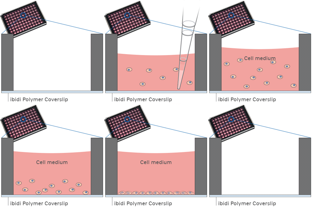
The Principle of the µ-Plate 96 Well Black
The Coverslip Bottom
The µ-Plate 96 Well Black comes with a thin ibidi Polymer Coverslip Bottom that has the highest optical quality (comparable to glass) and is ideally suitable for high-resolution microscopy. It is also available as a µ-Plate 96 Well Black Glass Bottom for special microscopic applications.
Find more information and technical details about the coverslip bottom of the ibidi chambers here.
The ibiTreat Surface
ibiTreat (tissue culture-treated) is our most recommended surface modification, because almost all adherent cells grow well on it without the need for any additional coating.
Find more information about the different surfaces of the ibidi chambers here.
Experimental Examples
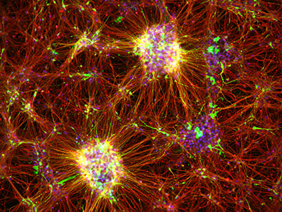
Immunofluorescence image of dopaminergic neurons derived from human induced pluripotent stem cells (iPSCs) in an ibidi μ-Plate 96 Well. The image shows the neurite extension with the expression of tyrosine hydroxylase (green), β-III Tubulin (red), and Foxa2 (blue). Images were acquired using a BZ-X710 microscope (Keyence). Image by Asuka Morizane, Kobe City Medical Center General Hospital, Kobe, Japan.
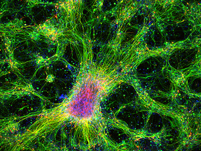
Immunofluorescence image of dopaminergic neurons derived from human induced pluripotent stem cells (iPSCs) in an ibidi μ-Plate 96 Well. The image shows the neurite extension with the expression of β-III Tubulin (green) and tyrosine hydroxylase (red). DAPI (blue) was used for nuclear staining. Image was acquired using a BZ-X710 microscope (Keyence). Image by Asuka Morizane, Kobe City Medical Center General Hospital, Kobe, Japan.
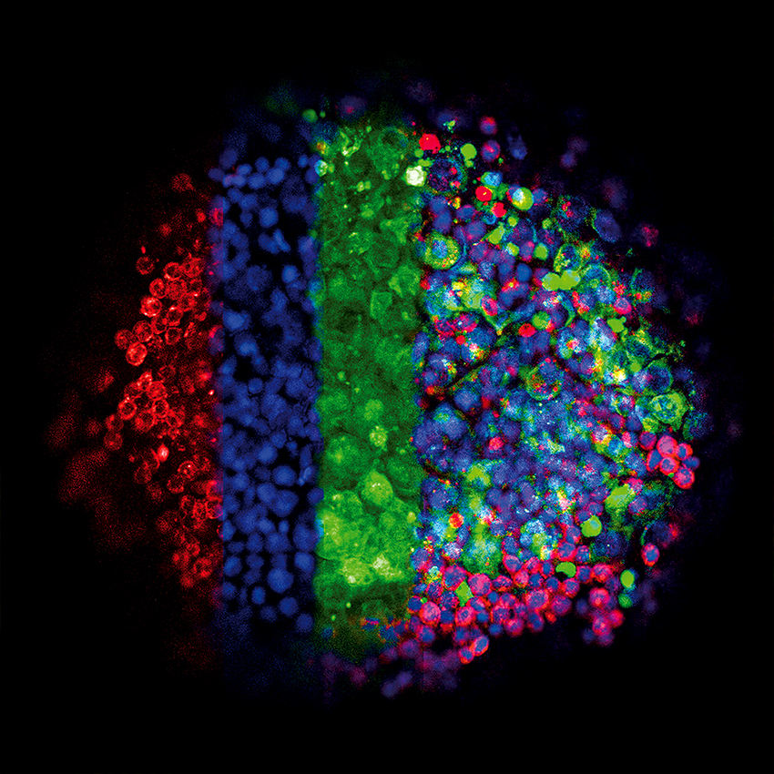
This image shows the three-dimensional growth of germ cell tumor cells (green, GFP) and immune cells (lymphocytes, red, mCherry) using the hanging drop technique. Cell nuclei appear in blue (DAPI). In 3D culture, a close interaction between the diverse cell types develops. The drops were transferred into an ibidi µ-Plate 96 Well. The image was acquired using a Zeiss LSM 710 confocal microscope with a 10x objective. Image by Gillian Ludwig and Daniel Nettersheim, Translational UroOncology, Department of Urology, University Hospital Düsseldorf, Germany.
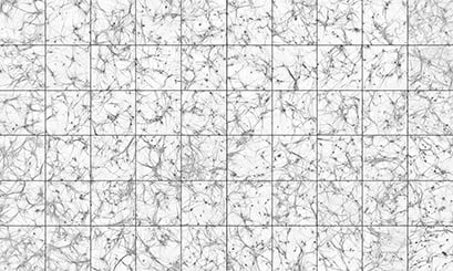
Human induced pluripotent stem cell (hiPSC) derived neurons stained with MAP2, a marker of the cell body and proximal dendrites. The neurons were differentiated via neurogenin 2 induction, and co-cultured with mouse glia for 30 days. Each square was recorded from a separate well of an ibidi µ-Plate 96 Well. The images were recorded on a high-content confocal imaging system with a 10x objective. Image by Chris Hempel, Q-State Biosciences, Inc., Cambridge, MA, USA.

High-content widefield fluorescence imaging of Rat1 fibroblast nuclei, stained with DAPI (blue) in the µ-Plate 96 Well Black. A complete scan of a single well contains 6 x 6 stitched microscope images (tile scanning mode), taken with a 10x objective lens.
