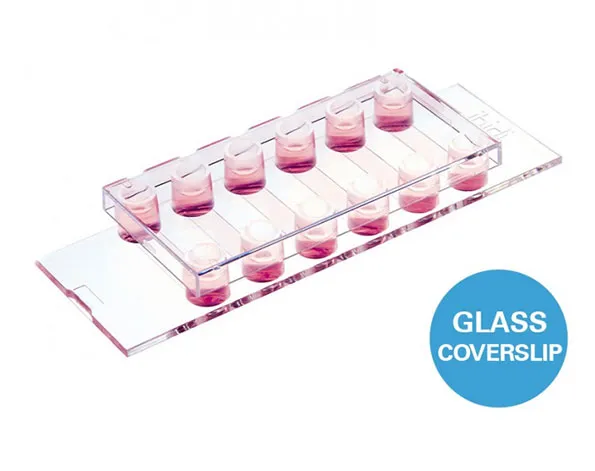
µ-Slide แบบ 6 ช่องสำหรับทำงาน flow assays และสำหรับ immonofluorescence
- สำหรับงาน immunofluorescence
- เซลล์กระจายตัวในช่องว่างอย่างทั่วถึง
- ประหยัดค่าใช้จ่ายในการทำการทดลองเนื่องมาจากใช้เซลล์จำนวนน้อย และใช้ reagent ในปริมาณน้อย
- สามารถใช้ในการถ่ายรูปได้ดีเนื่องมาจากก้นภาชนะทำมาจากแก้วที่มีความบางเท่ากับ coverslip (# 1.5H)
- มี 15 ชิ้น/ 1 กล่อง
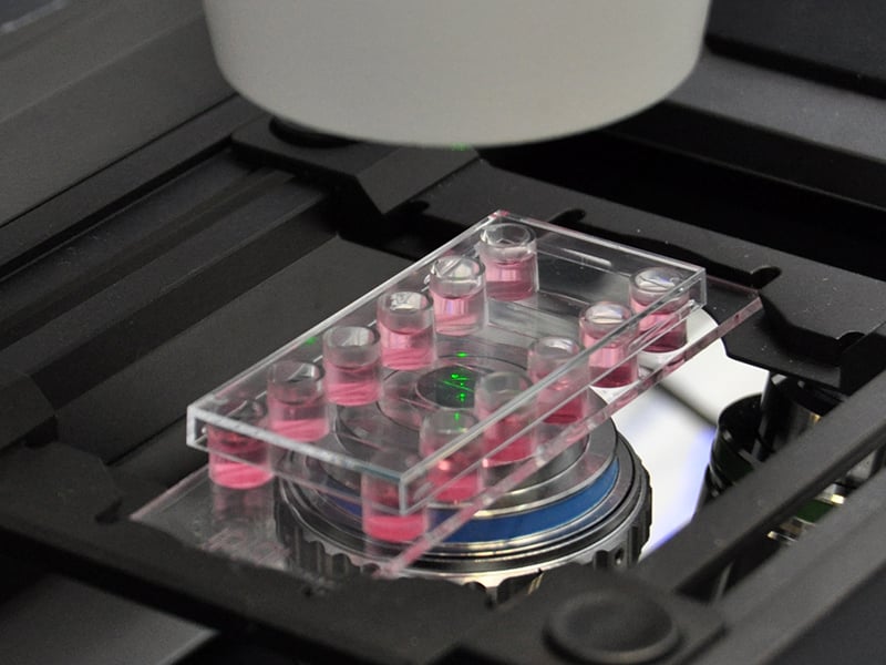
A 6 channel μ-Slide suitable for flow experiments and for immunofluorescence assays
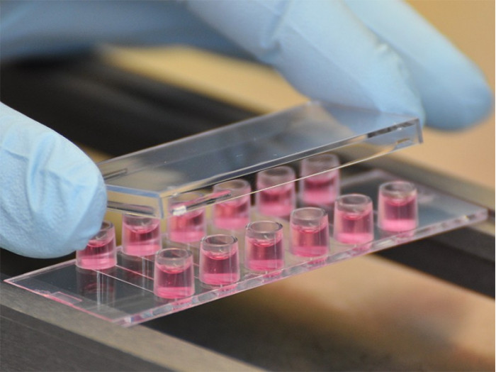
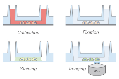
- All-in-one chamber that simplifies immunofluorescence protocols
- Homogeneous cell distribution over the channel surface, regardless of handling practices
- Cost-effective experiments with small numbers of cells and low volumes of reagents
- Perfect cell imaging thanks to the low thickness variability of the coverslips’ glass (# 1.5H)
- Surface Modification: #1.5H (170 µm +/- 5 µm) D 263 M Schott glass, sterilized
Pcs./Box: 15 (individually packed)
Applications
- Cultivation and high-resolution microscopy of cells
- TIRF and single molecule applications of living and fixed cells
- Super-resolution microscopy
- Immunofluorescence staining and fluorescence microscopy of living and fixed cells
- Live cell imaging
- Mounting immunofluorescent samples using the ibidi Mounting Medium and the ibidi Mounting Medium With DAPI
- Real-time imaging under either static or flow conditions
- Parallel screenings using multichannel pipettes
Specifications
| Outer dimensions (w x l) | 75.5 x 25.5 mm² |
| Adapters | Female Luer |
| Number of channels | 6 |
| Channel volume | 40 µl |
| Channel height | 0.54 mm |
| Channel length | 17 mm |
| Channel width | 3.8 mm |
| Volume per reservoir | 60 µl |
| Height with/without lid | 8.8 mm/7.6 mm |
| Growth area | 0.6 cm² per channel |
| Coating area using 40 µl | 1.2 cm² per channel |
| Bottom: Glass coverslip No. 1.5H, selected quality, 170 µm +/- 5 µm | |
Technical Features
- 40 µl channel volume, which saves on reagent consumption
- No. 1.5H High Precision D 236M borosilicate glass coverslip
- Fully compatible with high resolution fluorescence microscopy
- Easy connection to existing tubes and pumps via female Luer adapter
- Defined shear stress and shear rate level
- Also available as a µ-Slide VI 0.4 with a Polymer Coverslip Bottom

All-in-One-Chamber for Fast Immunofluorescence Protocols
Application Example: PAINT Super-Resolution Microscopy of B Cells
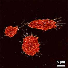
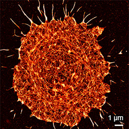
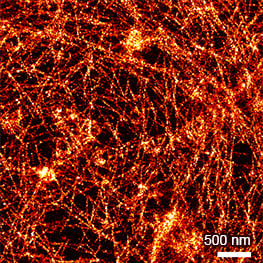
Super-resolution PAINT microscopy of fixed B cells in the µ-Slide VI 0.5 Glass Bottom, using a TIRF microscope. LifeAct-Cy3B was used to visualize the F-actin cytoskeleton. Objective lens: Nikon CFI SR HP Apochromat TIRF 100x Oil immersion, NA=1.49. Courtesy of Massive Photonics GmbH (www.massive-photonics.com).
ibidi Polymer Coverslip vs. ibidi Glass Bottom
ibidi Polymer Coverslip | ibidi Glass Bottom | |
| Optical properties | ||
| Refractive index (nD 589 nm) | 1.52 | 1.52 |
| Abbe number | 56 | 55 |
| Thickness | #1.5 (180 µm) | #1.5H (170 µm) |
| Material | Microscopy plastic | D 263M Schott borosilicate glass |
| Autofluorescence | Low | Low |
| Transmission | Very high (even ultraviolet) | High (ultraviolet restrictions) |
| Birefringence (DIC) | Low (DIC compatible*) | Low (DIC compatible*) |
| Other aspects | ||
| Surface modifications | ibiTreat – tissue culture treated Uncoated – hydrophobic | Only glass |
| Protein coatings | Compatible | Compatible |
| Gas permeable | Yes | No |
| Material flexibility | High | Low |
| Breakable | No | Yes |
| Applications | Fluorescence microscopy | TIRF and single photon |
