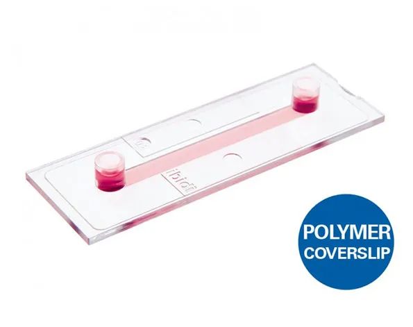
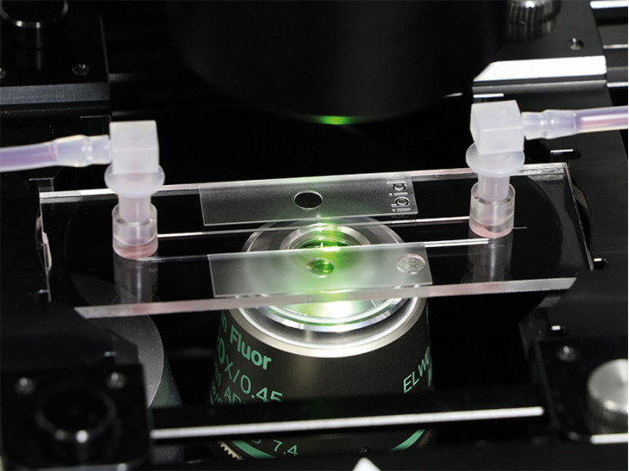
Channel slides โดยมีความสูง ปริมาตร และการเคลือบที่แตกต่างกัน – เหมาะสำหรับ flow applications
- พื้นที่ขนาดใหญ่ โดยมี shear stress สม่ำเสมอ
- มีความสูงให้เลือก 0.2, 0.4, 0.6 และ 0.8 มม.
- มี 15 ชิ้น/ 1 กล่อง
- เชื่อมต่อได้ง่ายโดยใช้ Luer adapters
- เซลล์กระจายตัวในเส้นทางที่กำหนดไว้
Channel slides with different heights, volumes, and coatings specially suited for flow applications
- Large area of uniform shear stress
- Easy connection using Luer adapters
- Homogeneous cell distribution with defined optical pathway
Applications
- Adherent cells under flow conditions
- Cell culture (static or stop-flow)
- Simulation of blood vessels with endothelial cells
- High-resolution microscopy of living and fixed cells
Specifications
| Outer dimensions (w x l) | 25.5 x 75.5 mm² |
| Channel length | 50 mm |
| Channel width | 5 mm |
| Adapters | Female Luer |
| Volume per reservoir | 60 μl |
| Growth area | 2.5 cm2 |
| Coating area | 5.2 / 5.4 / 5.6 / 5.8 cm2 |
| Bottom: ibidi Polymer Coverslip | |
Technical Features
- Standard format with thin bottom for low or high magnification microscopy (up to 100x)
- Large observation area for microscopy
- Channel volumes of 50 μl, 100 μl, 150 μl, or 200 μl
- Defined shear stress and shear rate levels
- Easy connection to tubes and pumps using Luer adapters
- Available as variety pack containing all heights
- Fully compatible with the ibidi Pump System
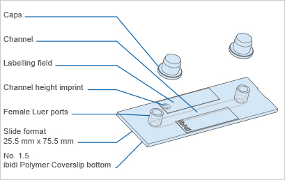
The Principle of the µ-Slide I Luer
The Coverslip Bottom
The µ-Slide I Luer comes with a thin ibidi Polymer Coverslip Bottom that has the highest optical quality (comparable to glass) and is ideally suitable for cell culture under flow and high-resolution microscopy. It is available with different channel heights or as a sticky version without any bottom.
Find more information and technical details about the coverslip bottom of the ibidi chambers here.
The ibiTreat Surface
ibiTreat (tissue culture-treated) is our most recommended surface modification, because almost all adherent cells grow well on it without the need for any additional coating.
Find more information about the different surfaces of the ibidi chambers here.
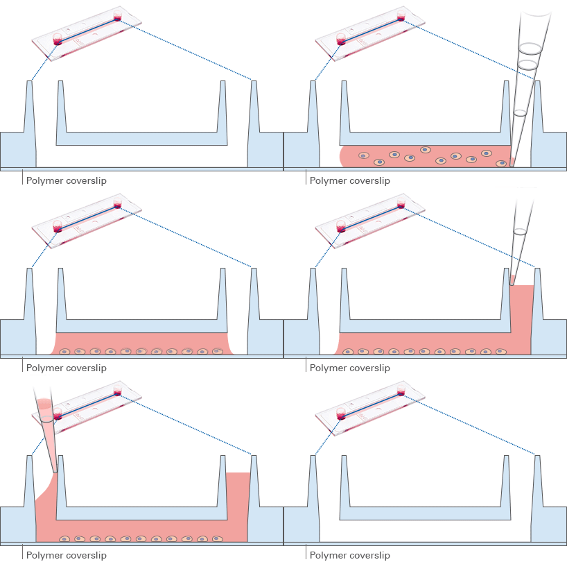
Choosing the Right Channel Height for Flow Applications
Which Experiment Are You Planning?
Low channels are more suitable for flow applications.
High channels are more suitable for static cell culture.
| For flow assays with small amounts of medium and high values of shear stress | 0.2 channel |
| For a wide range of shear stress | 0.4 channel |
| For controlling low values of shear stress < 2 dyne/cm2 | 0.6 and 0.8 channels |
Select the ideal Perfusion Set for your flow application here.
Cross Section of the Different Channel Heights

* not recommended for static culture over more than 6 hours
Experimental Examples
Cells Cultured Under Static or Flow Conditions
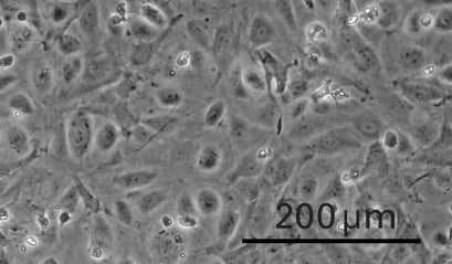
Static cultivation
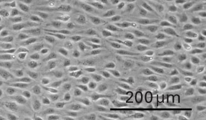
Flow cultivation
Influence of Shear Stress on Cultured Cells
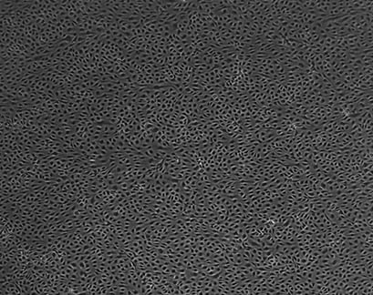
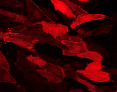
Human umbilical vein endothelial cells (HUVEC) cultured under flow conditions (20 dyn / cm²) in a µ-Slide I 0.4 Luer over 9 days. The primary cells were transduced with the adenoviral vector rAV CMV-LifeAct-TagRFP 24 hours prior to the experiment.
Which Slide Should I Use for My Application?
| µ-Slide Spheroid Perfusion | µ-Slide III 3D Perfusion | µ-Slide I Luer 3D | µ-Slide I Luer | |
| Application | ||||
| Perfusion of samples | Yes | Yes | Yes | Yes |
| Defined shear stress on cell monolayers | No | No | Yes, on gel matrix | Yes, on coverslip |
| Gel matrices for 3D | No | Yes | Yes | No |
| Cell Type | ||||
| Spheroids/organoids | Yes, free floating in well | Yes, inside gel matrix only | Yes, inside gel matrix only | No |
| Suspension cells | Yes, free floating in well | Yes, inside gel matrix only | Yes, inside gel matrix only | Yes, in flow suspension only |
| Adherent cells | Yes, on coverslip | Yes, inside or on gel matrix | Yes, inside or on gel matrix | Yes, on coverslip |
