Proven Technology for Diagnostic Excellence
เทคโนโลยีที่พิสูจน์แล้วเพื่อความเป็นเลิศด้านการวินิจฉัย
เทคโนโลยีที่ได้รับการพิสูจน์แล้วของ Aperio GT 450 DX มุ่งเน้นไปที่การเพิ่มประสิทธิภาพการทำงานของแล็บและการดูแลผู้ป่วย ซึ่งจะมีบทบาทสำคัญในการผลักดันการใช้งานพยาธิวิทยาดิจิทัลให้แพร่หลายยิ่งขึ้น ท่ามกลางข้อกำหนดด้านการควบคุมที่เข้มงวดมากขึ้น ผู้ผลิตอุปกรณ์พยาธิวิทยาดิจิทัลจึงต้องปฏิบัติตามมาตรฐานสูงสุดในด้านคุณภาพ ความปลอดภัยของข้อมูล และความปลอดภัยของผู้ป่วย
ไฮไลต์ของ Aperio GT 450 DX
- ภาพสแกนคุณภาพสูงแบบสม่ำเสมอ
ใช้เทคโนโลยีปรับเทียบสีอัตโนมัติ (color calibration) เพื่อให้ได้ภาพที่มีคุณภาพสูงอย่างสม่ำเสมอ ช่วยให้สามารถวินิจฉัยได้ตรงเวลา บนหน้าจอ - ออกแบบโดยคำนึงถึงพยาธิแพทย์
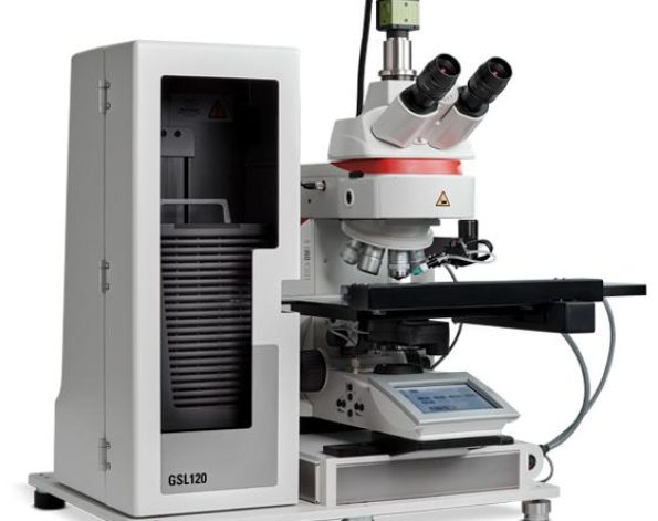
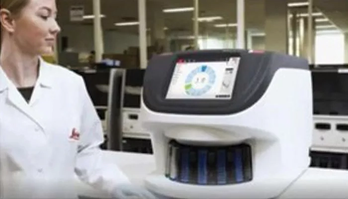
เป็นเครื่องสแกนสไลด์เนื้อเยื่อแบบดิจิทัลที่ ทำงานอัตโนมัติ, รองรับปริมาณงานสูง, และให้ คุณภาพภาพยอดเยี่ยม พร้อมการทำงานที่รวดเร็วและการดูภาพที่ลื่นไหล - ลดเวลา turnaround ของเคส
ด้วยฟีเจอร์สแกนอัตโนมัติแบบไม่ต้องสัมผัส (no-touch scanning) และการโหลดรางสไลด์ได้อย่างต่อเนื่องโดยตรงจากเครื่อง HistoCore SPECTRA Workstation (เครื่องย้อมและปิดแผ่นสไลด์) - ความปลอดภัยด้าน IT สูงขึ้น
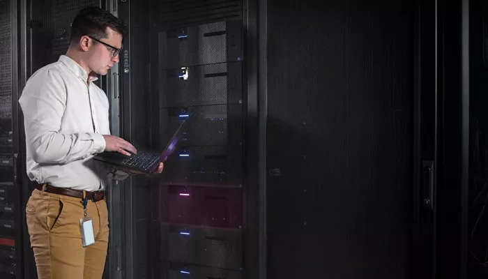
ด้วยระบบจัดการเครื่องสแกนแบบรวมศูนย์ผ่านเซิร์ฟเวอร์และซอฟต์แวร์ Scanner Administrator Manager DX (SAM DX) ที่ช่วยให้คุณสามารถควบคุมและตรวจสอบเครื่อง GT 450 DX หลายเครื่องได้ในเวลาเดียวกัน
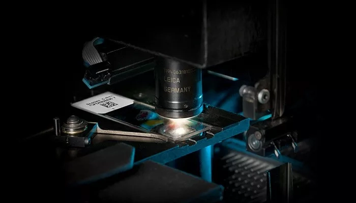
(ไม่จำเป็นต้องมีเวิร์กสเตชันแยกสำหรับแต่ละเครื่องอีกต่อไป) - ภาพคมชัดด้วยเลนส์ Leica optics
รับประกันคุณภาพของภาพด้วยเทคโนโลยีเลนส์จาก Leica ที่มีชื่อเสียงระดับโลกในด้านความแม่นยำและความคมชัด
Proven technology for improved workflow efficiency and patient care are set to define digital pathology and increase overall adoption. With the call for more rigorous controls, manufacturers of digital pathology devices are being held to the highest standards of quality, security, and patient safety.
The Aperio GT 450 DX uses innovative color calibration technology to output consistent high-quality images enabling on-time, on-screen diagnosis. Aperio GT 450 DX is an automated, high capacity digital pathology slide scanner that offers exceptional image quality, rapid delivery of cases and high-performance streamlined digital viewing built with the pathologist in mind.
- Improve case turnaround with no touch scanning and continuous loading of the racks directly from the HistoCore SPECTRA Workstation (Stainer & Coverslipper)
- Increase IT security and control with a dedicated Scanner Administrator Manager DX (SAM DX) server and software that allows you to set up and monitor multiple Aperio GT 450 DX scanners at a time. No more single workstations for each scanner
- Ensure excellent image quality with Leica optics
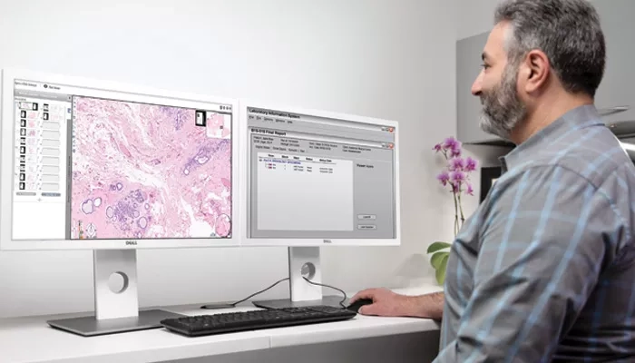
Confidence in Diagnosis for the Pathologist
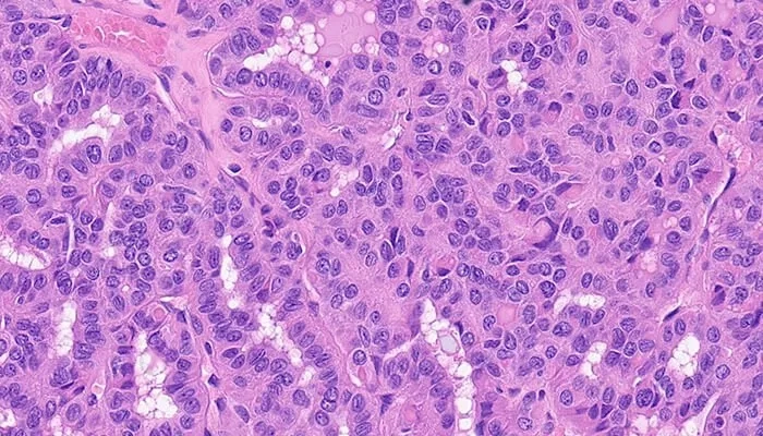
The Aperio GT 450 DX starts with quality and stays with quality. High quality digital scans deserve an image management system that detects, stores and delivers whole slide images and metadata exactly where and when needed for confidence in diagnosis.
Aperio GT 450 DX Scanner
- Designed to rapidly deliver images with exceptional quality for Pathologists.*
- High performance objective manufactured by Leica Microsystems, renowned for producing world-class optics since 1849.
- Leica optics deliver exceptional image quality
- Every slide is calibrated during scanning to ensure the best quality image
- Automated image quality assessment, so you don’t have to send slides back for re-scans 99.5% accurate tissue finder (H&E stains) finds faint tissue while excluding pen marks, dust, and residue.
Security and Interoperability for IT Professionals
The Aperio GT 450 DX system offers fast, secure, flexible IT architecture
Aperio GT 450 DX Scanner:
- HL7, and DICOM compatible for easy integration into LIS, LIMS or PACS
- A centralize SAM DX server and dedicated software provides you with a data management solution that can remotely set up and monitor multiple Aperio GT 450 DX scanners at a time.
- Comprehensive cybersecurity including encryption and login controls
- Centralize scanner management to set up and monitor multiple scanners
- Scalable set up for hub and spoke configurations
- Supports dedicated logins for each Aperio GT 450 DX scanner
*VP-0463 validation results
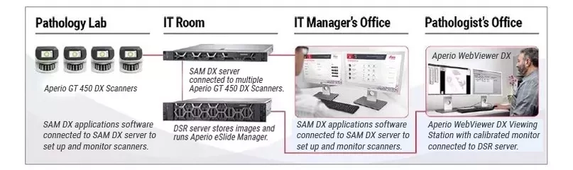
From the lab to the IT room to the office, the Aperio GT 450 DX was designed to scale up digital pathology operations for histotechnicians, IT professionals and pathologists.
Speed to Quality for Lab Managers and Histotechnicians
The Aperio GT 450 DX is simple to operate and helps lab managers and histotechnicians deliver rapid, quality results with confidence.
Aperio GT 450 DX scanner:
- No touch continuous loading during scanning
- Automated IQ checks during each scan to ensure quality
- Assign priority cases
- Each slide is calibrated individually prior to scan
- 450-slide capacity, packed into a small size, fits on the lab bench
- 99.5% accurate tissue finder (H&E Stains) allows you to adjust if needed*
- 100% first-time barcode scan success rate*
- Slides are then automatically scanned at 81 slides per hour at 40x for a 15mm x 15mm area.
- Fast delivery of urgent cases directly to the pathologist’s viewing station
Precision, Potential to Scale, and Peace of Mind
Digital pathology scan speeds have not always been fast enough at 40x magnification to keep up with high-volume scanning (120k+ slides per year). Real-Time Focusing (RTF)** offers a potential solution to this problem. It’s a novel method to capture a digital image that combines an imaging line sensor and a focusing line sensor.
- While scanning, the focusing line sensor receives focusing data of the tissue.
- Proprietary algorithms determine the best focus value.
- Then, each best focus value is treated like a control loop with the mechanics, feeding the best focus value to the objective height on the fly.
- The positioning adjustments of the objective are performed continuously (in real time) in order to enable the imaging line sensor to capture the image at its best possible focus. Hence, real time focusing.
Excellent Image Quality with Custom-Made Leica Objective
The high-performance objectives included with the Aperio GT 450 DX is specifically designed to maximise field of view for high speed digital pathology scanning. Most objectives are designed similarly to the human eye: round, which focuses light in the center and limits field of view. The objectives on the Aperio GT 450 DX includes an extra-wide flat field correction that enables a much larger field of view (1mm) that can accommodate extremely large digital images for fast scans and 0.26 um/pixel resolution at 40x magnification.
Product Specifications
IMAGING, CAPACITY & OPERATIONS
| Magnification: | 40x |
| Scan Speed: | 32 sec/slide (@ 40x for 15mm x 15mm area) |
| Scan Throughput: | 81 slides/hr (@40x for 15mm x 15mm area)* |
| Scan Resolution: | 0.26 µm/pixel at 40x |
| Scan Output: | DICOM compatible and SVS |
| Scanning Region: | ≤ 23.6 mm x 58 mm |
| Field of View (FOV): | 1mm |
| Objective Lens: | Custom-made by Leica Microsystems |
*VP-0450
| Slide Capacity: | 450 slides (15 racks of 30) |
| Slide Loading: | Automatic Continuous Load up to 450 1-inch x 3-in (2.54 cm x 7.62 cm) slides |
| Racks Accepted: | Leica Universal Rack (30-slide capacity), validated and recommended for use with HistoCore SPECTRA Workstation |
| Sakura Prisma Stainer and Coverslipper Rack (20-slide capacity) |
DIMENSIONS & WEIGHT
| Dimensions (W x L x H): | 52,83cm W, 71,12 cm D, 49,53cm H |
| Weight: | 63.5 kg (140 lbs) |
IT REQUIREMENTS & INTEGRATION
| Network Bandwidth Requirements (Aperio GT 450 DX to SAM Server): | 1 gigabits per second (Gbps) |
| Network Bandwidth Requirements (SAM Server to image repository/DSR): | 10 gigabits per second (Gbps) |
| Image File Formats: | DICOM compatible images, SVS, TIFF |
| Compatibility: | HL7 and DICOM (for LIS, LIMS, PACS integration) |
SECURITY FEATURES
| SSL for SAM User Interface | |
| Assign User Specific PINs per Scanner | –includes automatic timeouts |
| Secure Sign-On to SAM Server Software |
| Log Data & Events from Mirth Connect to File System | |
| Remote Access to Scanner Status | |
| Create User Groups and Assign Permissions | –password for client software and PIN for scanner |
SLIDES, COVERSLIPS & LABELS
| SLIDES | |
| General: | The tissue/smear is embedded between supported slide and coverslip |
| Standard: | 2.54 cm x 7.62 cm (1-inch x 3-inch) microscope slides. Measurements comply with ISO 8037/1. |
| Minimum Slide Size (W x L): | 25 mm x 75 mm |
| Maximum Slide Size (W x L): | 26 mm x 76 mm |
| Thickness: | optimized for range of 0.9 mm to 1.1 mm, excluding coverslip |
| Special Instructions: | The coverslip/label shall not protrude beyond the edge of the glass microscope slide. The entire coverslip and label must be glued to the glass microscope slide. There must be no lifted edges or parts of the coverslip/label. The outer surface of the slide must be dry. No liquid shall flow out of the specimen area. |
| Slide Preparation: | Slides are prepared typically using:Glass coverslip with mounting media, EukittFilm coverslip with integrated glue |
| Maximum Tissue/Smear Thickness: | optimised for tissue thickness of 3-5 μm (including mounting media) |
| COVERSLIPS | |
|---|---|
| Coverslips Accepted: | optimised for coverslip with thickness of .17 mm, made of typical coverslip material: Standard microscope cover glass or Cellulose Tri-Acetate film (microscope cover film). |
| LABELS | |
|---|---|
| Label Area (W x L): | 25 mm x 25 mm. Handwritten/printed non-transparent, matt (paper-like reflecting) sticker. |
| Label Special Instructions: | Labels shall not protrude beyond the edge of the slides or be lifted. Labels shall not be attached to the bottom of the slide, but only attached to the coverslip-side of the slide. |
| BARCODES | |
|---|---|
| Barcodes Engines: | 1D and 2D Symbology |
| Barcodes Supported: | NW7, QR Code, Data Matrix, Interleaved 2 of 5, Code 39, Code 128, PDF417, MicroPDF417 |