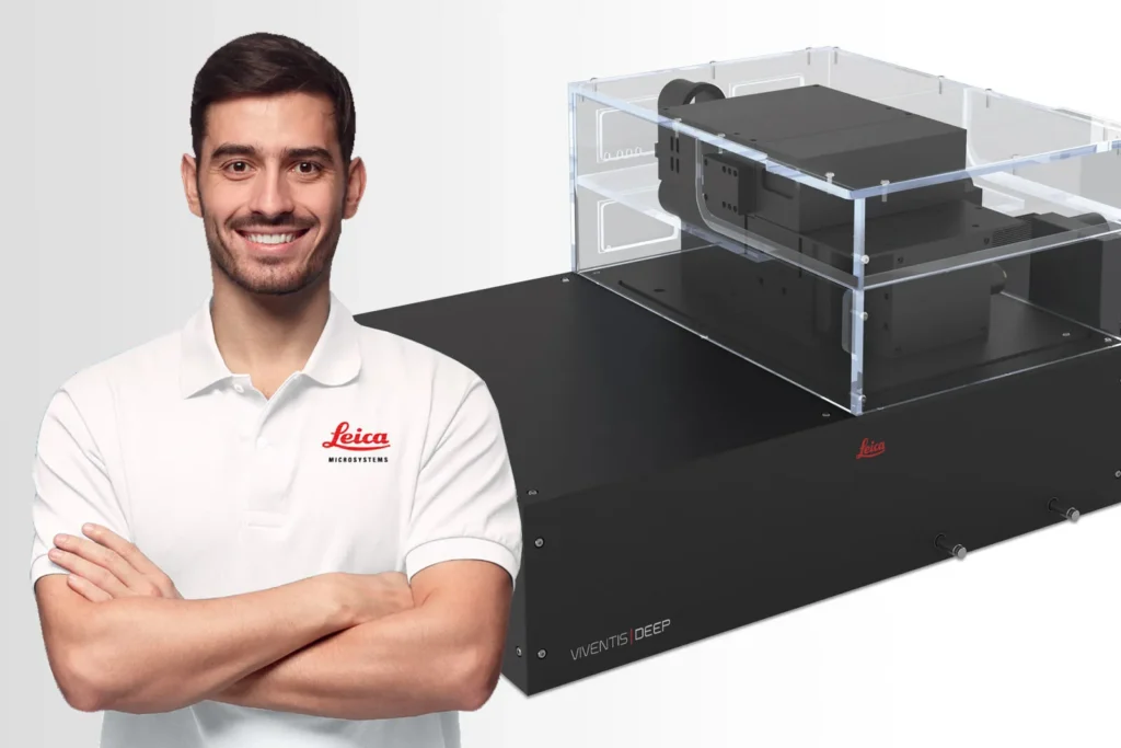
The Viventis Deep light sheet fluorescence microscope combines multi-view and multi-position light-sheet imaging to illuminate life in its entirety.
Begin your journey to discover deep, long-term imaging that reveals the intricate details and dynamics of biological systems.
Leica Viventis Deep เป็นกล้องจุลทรรศน์ฟลูออเรสเซนซ์แบบ Light Sheet ที่ออกแบบมาเพื่อการถ่ายภาพตัวอย่างชีวภาพที่มีความลึกและรายละเอียดสูง ด้วยเทคโนโลยีการส่องสว่างและการตรวจจับแบบคู่ (Dual View) รวมถึงความสามารถในการปรับตำแหน่งหลายจุด (Multi-Position) ช่วยให้การศึกษาระบบชีวภาพมีความละเอียดและครบถ้วนมากขึ้น
คุณสมบัติเด่น:
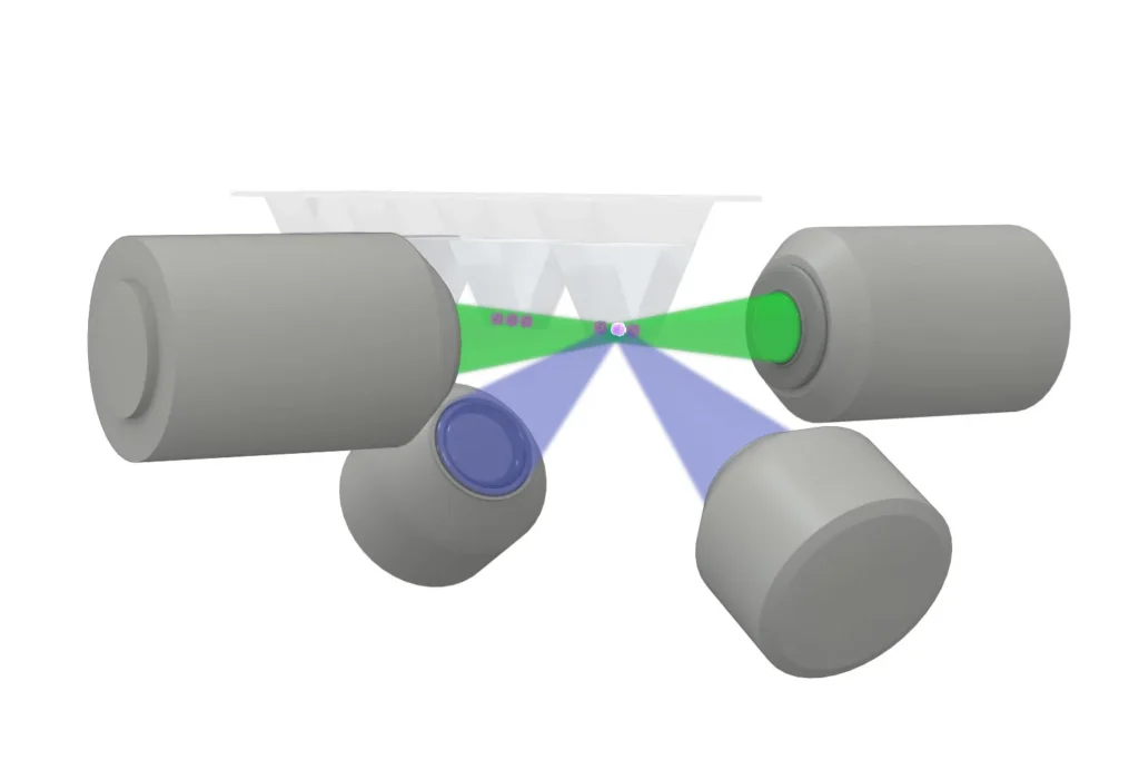
- การส่องสว่างและการตรวจจับแบบคู่: ใช้เลนส์ส่องสว่างขนาด 10x NA 0.2 สองตัว และเลนส์ตรวจจับขนาด 25x NA 1.1 หรือ 16x NA 0.8 สองตัว เพื่อเพิ่มความคมชัดและรายละเอียดในการถ่ายภาพ
- แหล่งกำเนิดแสงเลเซอร์หลายความยาวคลื่น: รองรับเลเซอร์สูงสุด 6 ความยาวคลื่น (จาก 405 nm ถึง 685 nm) เพื่อความยืดหยุ่นในการใช้งานและการปรับแต่งตามความต้องการ
- ความหนาของแสงชีตที่ปรับได้: มีแสงชีตที่ปรับความหนาได้สามค่า (ประมาณ 2.3, 4 และ 7 μm) เพื่อให้เหมาะสมกับขนาดและลักษณะของตัวอย่างที่แตกต่างกัน
- การจัดวางตัวอย่างที่สะดวก: ออกแบบด้วยช่องใส่ตัวอย่างด้านบนที่เปิดโล่ง (Open-Top) เพื่อความสะดวกในการติดตั้งและปรับเปลี่ยนตัวอย่าง รวมถึงการแลกเปลี่ยนสื่อเลี้ยงเซลล์ระหว่างการถ่ายภาพ
- การบำรุงรักษาสภาพตัวอย่าง: ระบบควบคุมอุณหภูมิและความเข้มข้นของ CO₂ และ O₂ ช่วยรักษาสภาพแวดล้อมที่เหมาะสมสำหรับตัวอย่างระหว่างการถ่ายภาพ
ด้วยคุณสมบัติเหล่านี้ Leica Viventis Deep จึงเป็นเครื่องมือที่เหมาะสำหรับการศึกษาระบบชีวภาพที่ต้องการความละเอียดสูงและการรักษาสภาพตัวอย่างที่ดี
Explore life in depth
The Viventis Deep microscope helps you to expand the spatio-temporal understanding of your sample to its full depth, thanks to increased spatio-temporal resolution.
Achieve detailed volumetric imaging for a complete view of the sample with a patented combination of
- Dual illumination
- Dual view detection
- Multi-position
- Open top sample holder
You can even image large light scattering samples over time with outstanding quality for meaningful downstream analysis, while minimizing light dose and maintaining sample accessibility.
Explore multiple living samples in parallel
Get more data from a single experiment. Collect data from multiple samples in parallel under different conditions in a single timelapse. The open-top configuration of the Viventis Deep microscope transforms your light sheet imaging with higher throughput and multi-position capabilities.
It enables
- Easy sample mounting
- Imaging under physiological conditions with only minor protocol changes
- Media exchange, even during a running time-lapse
Explore life events with long-term imaging
In a variety of model systems, including intestine, liver and human colon cancer organoids and zebrafish embryos, the gentle light sheet technology of the Viventis Deep microscope provides high image quality while preserving sample viability.
In addition, the advanced incubation solution and easy media exchange even during a running experiment preserves physiological conditions. Easily handle even more complex experimental conditions. To study responses and to ensure specific results, you can add drugs and use an optional photomanipulation arm during a running time-lapse.
Shape the light sheet technology to fit your needs
The Viventis Deep microscope helps you to tune the thickness of the scanned Gaussian beam (DLSM). You can tailor the light sheet with a focus on resolution or field of view.
Its flexible software allows you to change settings while the timelapse is running. In addition, a Python application programming interface (API) lets you code and plug in custom macros for your own experimental ideas.
To prevent your sample from moving out of the field of view (FOV) during time-lapse, intelligent online object tracking changes the stage coordinates.
Enabling your success with complete workflow support
Keep your operations running around the globe with best-in-class services entirely dedicated to microscopy and over 175 years of history.
Key features
- Leica Team: 500+ Service & Application experts
- Leica Training: 4-level factory certification program
- Leica Logistics: 5 regional hubs for genuine parts
- Leica OneCall: PhD-level hotline assistance
Viventis Deep Dual View Light Sheet Fluorescence Microscope
Revealing life in its full context
| Detection and Illumination optics | Illumination: two 10X objectives, NA 0.2.Detection: two 25X NA 1.1 OR two 16X NA 0.8 water immersion objectives. |
| Illumination | External laser combiner with a maximum of six wavelengths (from 405nm to 685nm), Omicron LightHUB+.Transmitted light illumination (LED light source) to locate the sample and acquire transmitted images. |
| Light sheet specification | Light sheet generated by scanning of a Gaussian beam.Three switchable light-sheets with thicknesses (FWHM) of approximately 2.3, 4 and 7 μm for different sample sizes.Automatic position-specific light-sheet alignment. |
| Detection camera | Two high sensitivity Hamamatsu ORCA-Fusion USB3 cameras.Field of view: 16X Objective: 536µm; 25X Objective: 599µmPixel size: 16X Objective: 406nm; 25X Objective: 260nm |
| Sample part | Motorized XYZ sample stage with maximum X travel range of 50mmMultiple sample located in an open-top FEP sample chamber (multi-well). |
| Sample incubation | Recirculating air temperature controller from 5°C above ambient temperature to 40°C.CO2 concentration control range 2-10%.O2 concentration control range 5-20%. |