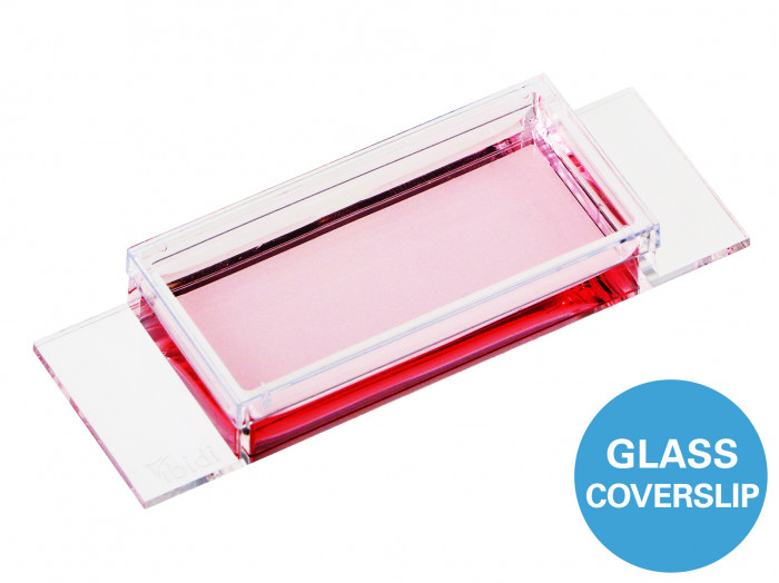
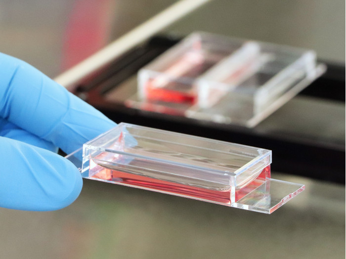
Chamber slide สำหรับเลี้ยงเซลล์ ขนาดความกว้าง 1 หลุม โดยมีพื้นสไลด์ทำจาก แก้ว Glass Coverslip ความหนา #1.5H สำหรับงานการถ่ายภาพแบบ TIRF และ Single molecule
A chambered coverslip with 1 large well and a #1.5H glass coverslip bottom for TIRF and single molecule applications
- An all-in-one cell culture chamber for cell culture and high-end imaging through the Glass Coverslip bottom
- Brilliant cell imaging thanks to the low thickness variability and highest optical quality of the Glass Coverslip bottom
- Maximal growth area for multiple applications: from culturing adherent cells to bioprinting
- Surface Modification: #1.5H (170 µm +/- 5 µm) D 263 M Schott glass, sterilized
Pcs./Box: 15 (individually packed) – 82107
Applications
- Cultivation and high-resolution microscopy of cells and tissues
- Flexible cell culture assays—from adherent cell culture to bioprinting
- TIRF and single molecule applications of living and fixed cells
- Super-resolution microscopy
- Immunofluorescence staining and fluorescence microscopy of living and fixed cells
- Live cell imaging over extended time periods
- Transfection assays
- Various microscopy techniques (e.g., widefield fluorescence, confocal microscopy, two-photon microscopy, FRAP, FRET, FLIM, and LSFM)
- Differential interference contrast (DIC) microscopy when used with a DIC lid
Specifications
| Outer dimensions (w x l) | 25.5 x 75.5 mm² |
| Number of wells | 1 |
| Dimensions of well (w x l x h) | 21.8 x 48.8 x 9.3 mm³ |
| Volume per well | 3 ml |
| Total height with/without lid | 10.8/9.5 mm |
| Growth area per well | 10.6 cm² |
| Coating area per well | 14.6 cm² |
| Bottom: Glass coverslip No. 1.5H, selected quality, 170 µm +/- 5 µm | |
Technical Features
- Open µ-Slide with 1 large well and a non-removable Glass Coverslip bottom
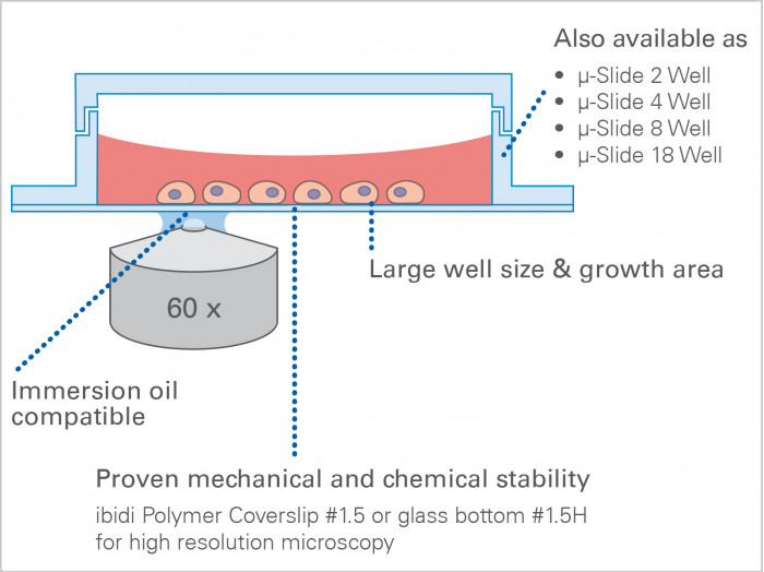
- Bottom made from D 263 M Schott glass, No. 1.5H (170 +/- 5 µm)
- May require coating to promote cell attachment
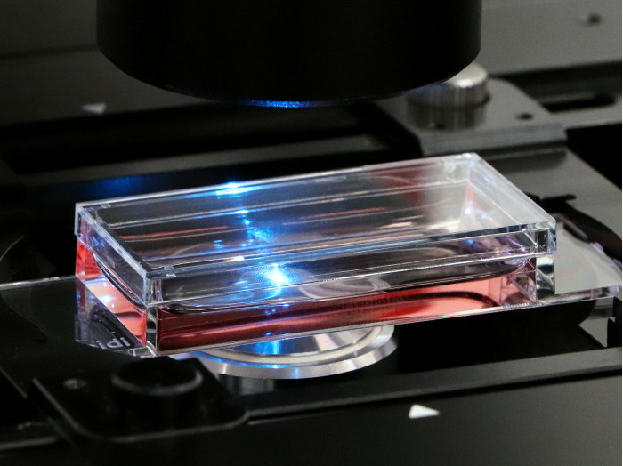
- Imaging chamber slide with excellent optical quality for high-end microscopy
- Closely fitting lid for low evaporation
- Compatible with staining and fixation solutions
- Made of fully biocompatible materials
- Also available with an ibidi Polymer Coverslip Bottom for superior cell growth: µ-Slide 1 Well


Application Examples


Widefield fluorescence microscopy of immunostained Rat1 cells in a µ-Slide 1 Well Glass Bottom. The F-actin cytoskeleton was stained with phalloidin (green). α-Tubulin antibodies were used to stain the microtubules (red). Nuclei were visualized with DAPI (blue). The image was taken on a Nikon microscope using a 10x (left) and a 20x (right) objective lens, the latter with oil immersion.

The large well area is ideal for various applications such as bioprinting.

The coverslip bottom is ideal for high resolution microscopy.
