")
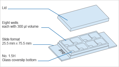
A chambered coverslip with 8 wells and a #1.5H glass bottom, suitable for use in TIRF and single molecule applications
- A cell culture chamber for observation of the sample through the coverslip-like bottom using high resolution microscopy
- A microscopy slide for brilliant cell imaging thanks to the low thickness variability of the coverslips’ glass
- Cost-effective experiments using small numbers of cells and low volumes of reagents
- Surface Modification: #1.5H (170 µm +/- 5 µm) D 263 M Schott glass, sterilized
Pcs./Box: 15 (individually packed) - Surface Modification: #1.5H (170 µm +/- 5 µm) D 263 M Schott glass, sterilized
Pcs./Box: 90 (individually packed)
Applications
- Cultivation and high-resolution microscopy of cells
- TIRF and single molecule applications of living and fixed cells
- Super-resolution microscopy
- Immunofluorescence staining and fluorescence microscopy of living and fixed cells
- Live cell imaging over extended time periods
- Transfection assays
- Differential interference contrast (DIC) microscopy when used with a DIC lid
Want to know if you should use a glass or a polymer bottom for your application? Find out here.
Specifications
| Outer dimensions (w x l) | 25.5 x 75.5 mm² |
| Number of wells | 8 |
| Dimensions of wells (w x l x h) | 9.4 x 10.7 x 6.8 mm³ |
| Volume per well | 300 µl |
| Total height with lid | 8 mm |
| Growth area per well | 1.0 cm² |
| Coating area per well | 2.20 cm² |
| Bottom: Glass coverslip No. 1.5H, selected quality, 170 µm +/- 5 µm | |
Technical Features
- Open µ-Slide with 8 independent wells
- Bottom made from D 263 M Schott glass, No. 1.5H (170 +/- 5 µm)
- May require coating to promote cell attachment
- Available as a Bulk Pack with 90 individually packed µ-Slides per box
- Also available as a µ-Slide 8 Well with an ibidi Polymer Coverslip Bottom for superior cell growth
- Now also available as a µ-Slide 8 Well high Glass Bottom with extra high individual walls to keep cross contamination between wells as low as possible
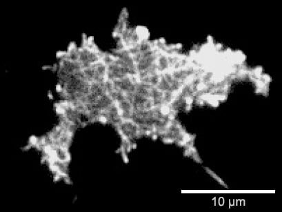
Experimental Example
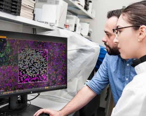
Surface-near F-actin network of a Dictyostelium discoideum DdLimE-GFP cell. Live cell imaging on a Glass Coverslip #1.5H using Total Interference Reflection Fluorescence (TIRF) microscopy.
ibidi Polymer Coverslip vs. ibidi Glass Bottom
ibidi Polymer Coverslip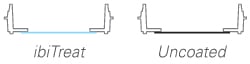 | ibidi Glass Bottom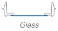 | |
| Optical properties | ||
| Refractive index (nD 589 nm) | 1.52 | 1.52 |
| Abbe number | 56 | 55 |
| Thickness | #1.5 (180 µm) | #1.5H (170 µm) |
| Material | Microscopy plastic | D 263M Schott borosilicate glass |
| Autofluorescence | Low | Low |
| Transmission | Very high (even ultraviolet) | High (ultraviolet restrictions) |
| Birefringence (DIC) | Low (DIC compatible*) | Low (DIC compatible*) |
| Other aspects | ||
| Surface modifications | ibiTreat – tissue culture treated Uncoated – hydrophobic | Only glass |
| Protein coatings | Compatible | Compatible |
| Gas permeable | Yes | No |
| Material flexibility | High | Low |
| Breakable | No | Yes |
| Applications | Fluorescence microscopy | TIRF and single photon |