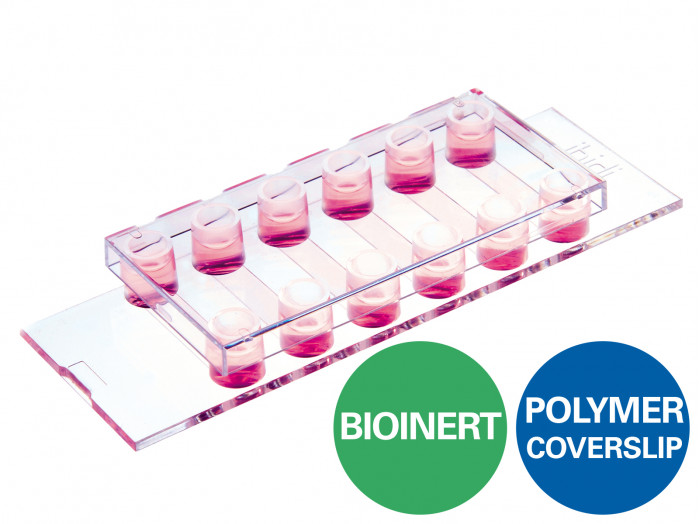
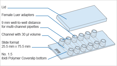
A 6 channel μ-Slide with a non-adherent surface for flow experiments and fluorescence microscopy
- Superior to the standard ultra-low attachment (ULA) surfaces—stable, biologically inert surface for long-term experiments without any cell or biomolecule adhesion
- Excellent microscopic quality without any autofluorescence—100% surface passivation combined with the highest optical quality of the ibidi Polymer Coverslip
- Pcs./Box: 15 (individually packed)
Surface Modification: Bioinert: #1.5 polymer coverslip, surface passivation with Bioinert, sterilized
Applications:
- Real-time imaging under either static or flow conditions
- Generation and long-term culture of 3D cell aggregates (e.g., spheroids and organoids) without any scaffold
- High resolution fluorescence microscopy of organoids, spheroids, embryoid bodies (EBs), and cells in suspension
- Background-free analysis of cell-cell interactions
- Prevention of stem cell differentiation due to attachment
- Culture of suspension cells in a permanently unattached state
- Self-assembly tumor spheroid formation assays
- 3D tumor spheroid models
Specifications
Bioinert Surface
| Bioinert surface thickness | 200 nm |
| Bioinert surface material | Polyol-based hydrogel |
| Protein coatings | Not possible |
µ-Slide VI 0.4
| Outer dimensions (w x l) | 75.5 x 25.5 mm² |
| Adapters | Female Luer |
| Number of channels | 6 |
| Channel volume | 30 µl |
| Channel height | 0.4 mm |
| Channel length | 17 mm |
| Channel width | 3.8 mm |
| Volume per reservoir | 60 µl |
| Growth area | 0.6 cm² per channel |
| Coating area using 30 µl | 1.2 cm² per channel |
| Bottom: ibidi Polymer Coverslip with Bioinert surface | |
Technical Features
- Channel µ-Slide with 6 independent channels and a non-removable polymer coverslip-bottom
- µ-Slide VI 0.4 with a non-adherent, passivated Bioinert surface
- Bioinert surface with long term passivation—superior to standard ULA surfaces:
- Ready-to-use without prior surface treatment or preparation
- No adsorption, coating, or binding of proteins, antibodies, enzymes, and other biomolecules
- Non-cytotoxic, biologically inert, and non-degradable
- Easy connection to existing tubes and pumps via female Luer adapters
- Imaging µ-Slide with excellent optical quality for high-end microscopy
- Compatible with staining and fixation solutions
- Biocompatible polymer material—no glue, no leaking
- Also available with the ibidi Polymer Coverslip bottom with a tissue culture-treated surface: µ-Slide VI 0.4 ibiTreat for various experiments with adherent cells
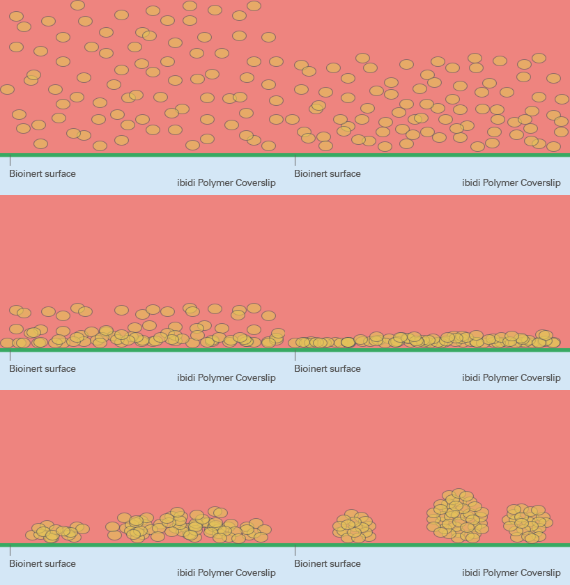
ibiTreat, Uncoated, and Bioinert—A Surface Comparison
ibiTreat
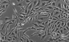
Excellent cell adhesion
- Culture of adherent cells
- ECM coatings possible
The hydrophilic ibiTreat surface provides excellent cell adhesion, even without a defined protein coating. However, ECM protein coatings can be done on ibiTreat without any restrictions. The ibiTreat surface is ideal for culture of adherent cells that do not need any specific coating.
Uncoated
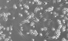
Weak cell adhesion
- Culture of adherent cells
- ECM coatings possible
The hydrophobic Uncoated surface provides weak cell adhesion, if not previously coated with an ECM protein. ECM protein coatings can be done on the Uncoated surface without any restrictions. The Uncoated surface is ideal for the culture of adherent cells that require a specific coating.
Bioinert
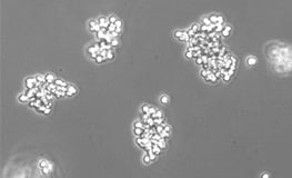
No cell adhesion
- Culture of suspension cells, cell aggregates, spheroids, embryoid bodies (EBs), organoids
- ECM coatings not possible
The hydrophilic Bioinert surface hinders any protein attachment, thus inhibiting subsequent cell attachment. Unlike with the ibiTreat and Uncoated surfaces, a coating is not possible. The Bioinert surface is ideal for the culture of suspension cells and cell aggregates.
