Linkam : CMS196V³
Linkam CMS196V³ เป็นระบบการถ่ายภาพแบบ Cryo-Correlative Light and Electron Microscopy (Cryo-CLEM) ที่ออกแบบมาเพื่อการศึกษาตัวอย่างที่อุณหภูมิต่ำมาก นี่คือคุณสมบัติหลัก:
- การรักษาตัวอย่างที่อุณหภูมิต่ำ: ใช้การทำความเย็นด้วยไนโตรเจนเหลวเพื่อรักษาตัวอย่างให้อยู่ในสภาพ vitrified ที่อุณหภูมิ -196°C2.
- การควบคุมอัตโนมัติ: มีระบบเติมไนโตรเจนเหลวอัตโนมัติที่ช่วยรักษาตัวอย่างให้อยู่ในสภาพ vitrified ตลอดเวลา.
- การเคลื่อนย้ายตัวอย่างที่แม่นยำ: มีแท่น XY ที่ควบคุมด้วยมอเตอร์และเข้ารหัสเพื่อการเคลื่อนย้ายตัวอย่างที่แม่นยำและการทำแผนที่พิกัดที่มีความละเอียดสูง3.
- การป้องกันการปนเปื้อน: ระบบการจัดการและการถ่ายโอนตัวอย่างที่ปลอดภัยและปราศจากการปนเปื้อน.
- การใช้งานร่วมกับกล้องจุลทรรศน์: รองรับการใช้งานร่วมกับกล้องจุลทรรศน์แบบตั้งตรงที่มีคุณภาพสูงจากผู้ผลิตหลายราย
CMS196V³ Cryo-CLEM Stage : overview
CMS196V³เป็นเครื่องมือ cryo-CLEM เพื่อเปิดใช้งานเวิร์กโฟลว์ทั้งหมดของ CLEM นอกเหนือจากการรักษาตัวอย่างที่ถูกทำให้เป็นผลึกโดยใช้การทำให้เย็นลงด้วยไนโตรเจนเหลวแล้ว ยังให้ความสามารถที่ได้รับการพิสูจน์แล้วว่าสามารถจัดการและถ่ายโอนตัวอย่างไครโอได้อย่างปลอดภัยและถ่ายภาพด้วยกล้องจุลทรรศน์แบบออปติคัลในขณะที่รักษาให้ปราศจากการปนเปื้อนตลอดเวลา รุ่นล่าสุดที่เข้ารหัสแบบใช้มอเตอร์และแบบใช้มอเตอร์ ร่วมกับซอฟต์แวร์ LINK ใหม่ช่วยให้สามารถผลิตแผนที่ที่มีความละเอียดสูงของกริดได้อย่างรวดเร็วและง่ายดาย นี่เป็นวิธีการตรงไปตรงมาของการทำแผนที่พิกัดที่จำเป็นในการค้นหาตัวอย่างเดียวกันในกล้องจุลทรรศน์เรืองแสงและใน EM
แม้ว่า CMS196V³ ได้รับการออกแบบมาโดยเฉพาะเพื่อแก้ปัญหาในการรับกริด EM ที่เป็นแก้วจากกล้องจุลทรรศน์เรืองแสงลงใน cryo TEM โดยไม่ต้องแยกส่วนและการปนเปื้อนผ่านการควบแน่น เวทียังต้องได้รับการปรับให้เหมาะสมทางแสงเพื่อให้สามารถใช้เลนส์ NA สูงได้
ใส่กริดสูงสุด 3 ช่องลงในตลับที่ออกแบบมาเป็นพิเศษซึ่งขนส่งจากช่องแช่แข็งแบบแช่ตัวในภาชนะขนาดเล็กที่ปิดสนิท บรรจุภาชนะลงในห้องบรรจุตัวอย่างแห้งที่ทำความเย็นไว้ล่วงหน้าซึ่งติดตั้งบนกล้องจุลทรรศน์เรืองแสงแบบตั้งตรง จากนั้นโหลดตลับเทปลงบนสะพานรับชมอย่างง่ายดายโดยใช้เครื่องมือปรับแต่งพิเศษ
คุณสมบัติ / จุดเด่นของ CMS196V³
- ระบบการสังเกตตัวอย่างด้วยความเย็นในตัวทำให้ตัวอย่างกลายเป็นแก้วที่อุณหภูมิ -196°C คงที่และหลีกเลี่ยงการปนเปื้อน
- เติมไนโตรเจนเหลวอัตโนมัติเติมของห้อง
- เวลาเริ่มต้นสั้นเพียง 10 นาที ไม่มีการเชื่อมต่อทางกลระหว่างตัวยึดตัวอย่างกับ Dewar ความเสถียรดีมาก ความเบี่ยงเบนต่ำในช่วงนาโนเมตร ความเสถียรในระยะยาวที่ดี
- แท่น XY แบบใช้มอเตอร์ในตัวพร้อมตัวเข้ารหัสแบบออปติคัลสำหรับการเคลื่อนไหวที่มีความแม่นยำสูงและการอ่านตำแหน่งที่ดีกว่า 1µm
- การทำแผนที่ XY แบบอัตโนมัติเต็มรูปแบบ
- ตัวเลือกการติดตั้งสำหรับกล้องจุลทรรศน์แบบตั้งตรงเกรดการวิจัยส่วนใหญ่จากผู้ผลิตรายใหญ่ทั้งหมด
- ระบบตลับตัวอย่างแม่เหล็กปรับแนวได้เองสำหรับตารางตัวอย่างสูงสุดสามตาราง
- รองรับ EM-grids, Auto-Grids (FEI), Membrane carriers (Leica HPM100), Bessy grids, planchettes, CryoCapsule จาก CryoCapCell และตัวยึด
- ตัวอย่างแบบกำหนดเอง ไม่จำเป็นต้องจัดการกับกริดเปล่า
- ตลับใส่ตัวอย่างสำหรับการโหลด จัดเก็บ และถ่ายโอนตัวอย่างที่ปลอดภัยและปราศจากการปนเปื้อน
- เชื่อมต่อกับโปรโตคอลการเตรียมตัวอย่างสำหรับการแช่แข็งแบบแช่ตัวและแช่แข็งด้วยแรงดันสูง
- เซ็นเซอร์ในตัวสำหรับการตรวจสอบอุณหภูมิตัวอย่าง
- ออปติกคอนเดนเซอร์ในตัวสำหรับช่องแสงที่ส่องผ่านและคอนทราสต์เฟส
- การสังเกตการเรืองแสงด้วย NA สูงถึง 0.9
- ทำงานอิสระเต็มวันด้วยตัวเลือกเติมอัตโนมัติ 3L LN2 Dewar
- จอแสดงผล LCD พร้อมปุ่มควบคุม การทำงานของจอยสติ๊กแบบสแตนด์อโลน และการควบคุมซอฟต์แวร์เต็มรูปแบบผ่านอินเทอร์เฟซ USB
- วงจร “อบออก” ทำความร้อนอัตโนมัติเพื่อไล่ความชื้น
- มากกว่า 50 ระบบที่พิสูจน์แล้วในภาคสนามทั่วโลก เป็นผู้นำตลาดใน Cryo-CLEM
Linkam : CMS196V³
Cryo-Correlative Microscopy Stage
Cryo-Correlative Microscopy Stage
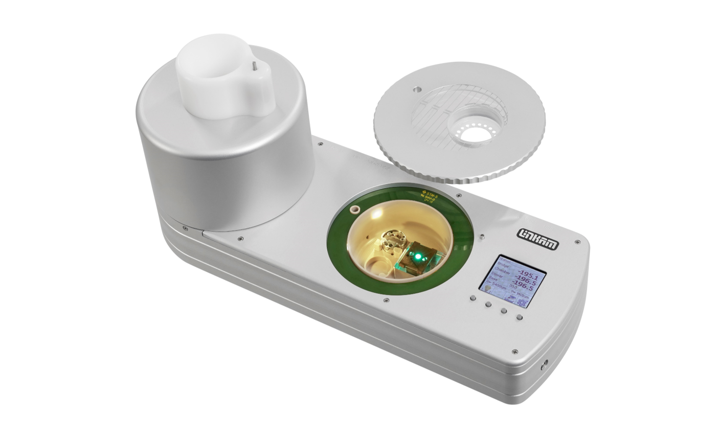
Developed in collaboration with Prof. Bram Koster’s team at Leiden University Medical Centre (LUMC) in the Netherlands, the CMS196 is the result of 4 years of R&D.
“The correlative stage was developed out of the necessity for a tool that uses fluorescence microscopy to help localise structures of interest in samples for high resolution cryo electron microscopy imaging. Over the years the system has evolved into an effective and efficient instrument that enables the imaging of cryo electron microscope images after light microscopy characterisation routinely.”
Prof. A.J. Koster – LUMC
CMS196V³ Cryo-CLEM Stage : overview
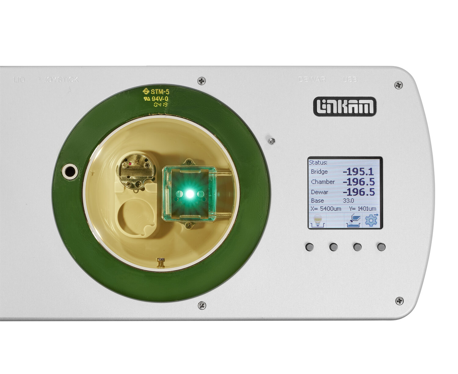
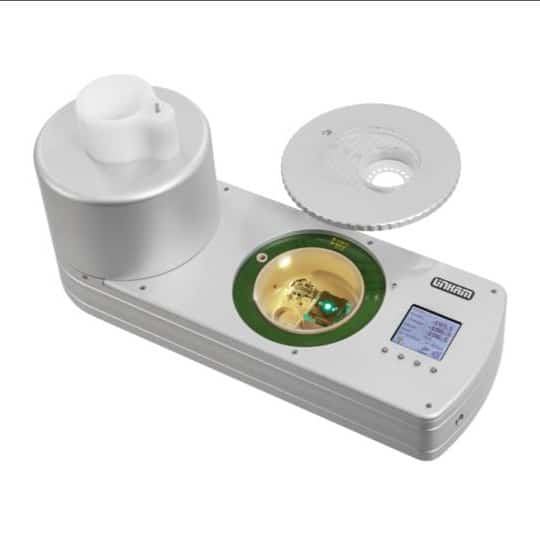
The CMS196V³ is a cryo-CLEM instrument to enable the full workflow of CLEM. Apart from maintaining the sample vitrified by means of liquid nitrogen cooling it provides proven capabilities to safely handle and transfer cryo samples and image them with optical microscopy while keeping them free of contamination at all times. The integrated motorised, encoded stage of the latest model, together with the new LINK software enables high resolution maps of the grid to be produced quickly and easily. This provides a straight forward method of coordinate mapping required to locate the same sample in the fluorescence microscope as well as in the EM.
Although the CMS196V³ was designed specifically to solve the problem of how to get vitrified EM grids from the fluorescent microscope into the cryo TEM without devitrification and contamination through condensation, the stage also had to be optimized optically to enable the use of high NA lenses.
Up to 3 grids are loaded into a specially designed cassette which is transported from a plunge freezer in a small sealed container. The container is loaded into a precooled dry sample loading chamber fitted on an upright fluorescent microscope. The cassette is then easily loaded onto the viewing bridge using special manipulation tools.
Features / Highlights of the CMS196V³
- Self-contained cryo sample observation system keeping the sample vitrified at constant -196°C and avoiding contamination
- Automatic top-up liquid nitrogen top up of the chamber
- Short startup time of only 10 minutes, no mechanical connection between sample mount and Dewar, very good stability, low drift in nm range, good long-term stability
- Integrated motorised XY stage with optical encoders for high precision movement and position readout of better than 1µm
- Fully Automated XY Mapping
- Mounting option for most research grade, upright microscopes from all major manufacturers
- Self-aligning magnetic sample cassette system for up to three sample grids
- Support for EM-grids, Auto-Grids (FEI), Membrane carriers (Leica HPM100), Bessy grids, planchettes, CryoCapsule from CryoCapCell and custom sample holders, no need to handle bare grids
- Sample cassette holder for safe and contamination-free sample loading, storage and transfer
- Connects to plunge-freezing and High-Pressure Freezing sample preparation protocols
- Integrated sensors for sample temperature monitoring
- Integrated condenser optics for transmitted light brightfield and phase contrast
- Fluorescence observation with NA of up to 0.9
- Full day of independent operation with optional 3L autofill LN2 Dewar
- LCD display with button control, standalone joystick operation and full software control via USB interface
- Automated heating “bake-out” cycle to drive out moisture
- With over 100 systems now in the field around the world, the CMS196 is the proven market leader in Cryo-CLEM
NEED MORE INFORMATION?
Contact us and one of our technical experts will be in touch shortly.
CMS196M automated mapping of an EM grid at high resolution
Why Cryo-CLEM?
Electron microscopy (EM) provides structural information at very high resolution. However, it can give only restricted insight into biological and chemical processes due to limitations in staining and sample preparation processes. Fluorescence microscopy on the other hand is a very sensitive method to detect biological, chemical and genetic processes and events inside living cells.
Cryo-CLEM brings it all together: it is a new and emerging technique that combines the individual advantages from both Fluorescence and EM by imaging the same sample location with both techniques and superimposing the complementary information.
“We are using the cryo-correlative stage to screen cells trapped in a thin layer of ice prior to imaging the samples at synchrotrons in Oxford, Barcelona and Berlin. The team at Linkam Scientific have helped to drive forward our research, quickly adapting and refining the technology to deliver an elegant, flexible, user-friendly stage.”
Dr. Lucy Collinson -Head of Electron Microscopy, The Francis Crick Institute
Imaging of vitrified samples under cryo-conditions
In order to make a biological sample compatible with the vacuum conditions found in EM and preserve the structural detail, samples are embedded in vitrified “glass-like” ice and need to be kept below -140°C. Any contact with moisture contained in the air has to be avoided since ice crystals would form immediately and contaminate the sample. Under cryo-conditions the fluorescence signals’ structural detail is preserved and photobleaching is significantly reduced.
Read about the new Linkam CryoGenium plunge freezing system (under development)
Linkam Co-ordinate transform tool
