RWD : RFLSI Ⅲ Laser Speckle Imaging System – RFLSI Ⅲ ระบบสร้างภาพด้วยแสงเลเซอร์
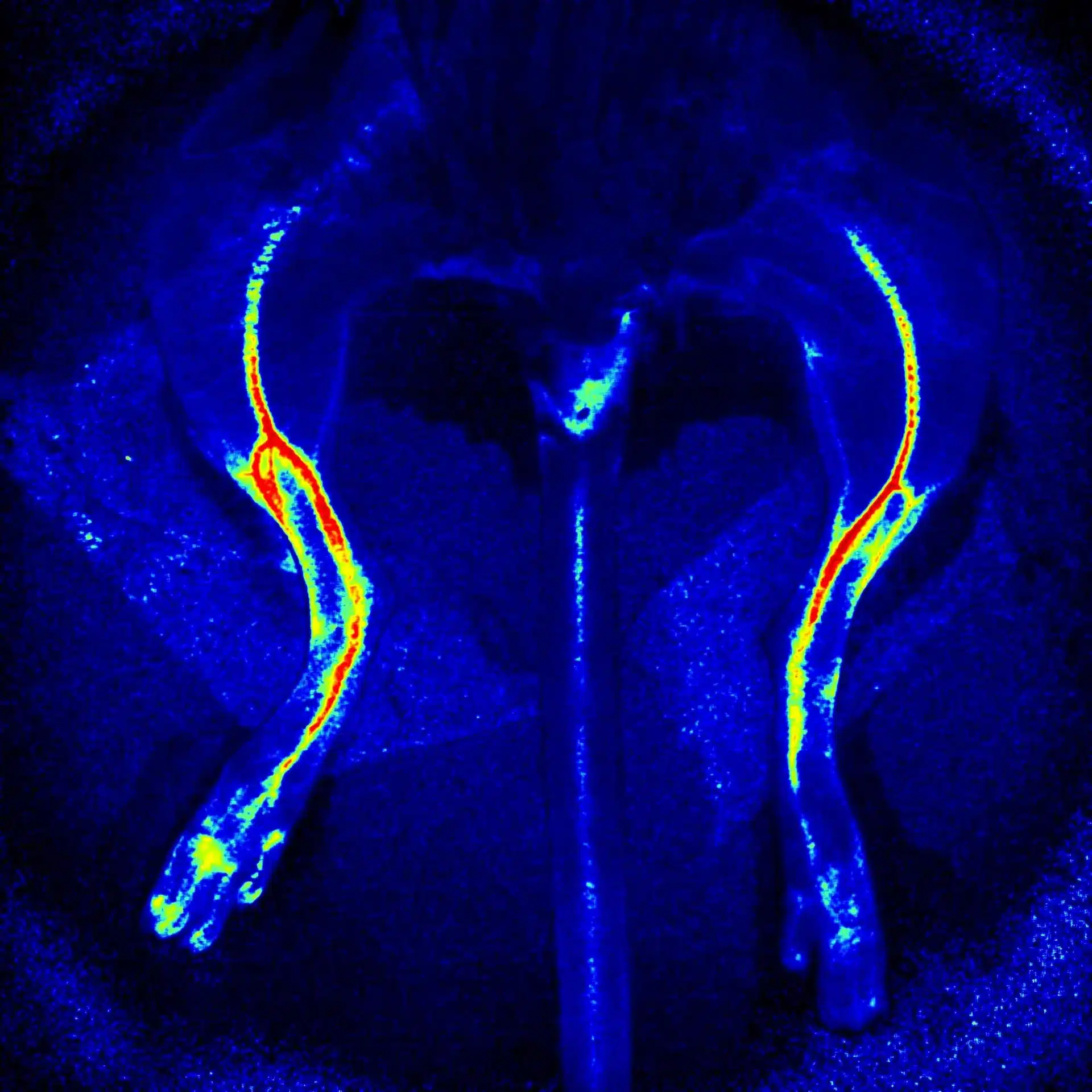
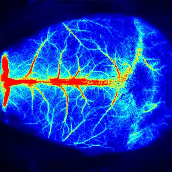
RFLSI III is a robust blood perfusion imaging system. Along with advanced software, it helps researchers real-time monitor and record blood perfusion of any exposed tissues or organs for microcirculation study, to visualize the quantified data directly, shorten the experiment time, and obtain outcomes easily.
Overview
RFLSI Ⅲ is based on the LSCI (Laser Speckle Contrast Imaging) technology. With the advantages of its non-contact, high frame-rate, high spatial resolution, it can be used to observe and record blood perfusion of any exposed tissues or organs for microcirculation study.
This system is designed for pre-clinical microcirculation researches like ischemic stroke, lower limbs, mesentery, etc. Multi-Output includes blood perfusion images and videos (4.2 million pixels), quantified data for perfusion unit and vessel diameter.
Applications:
- Cerebral blood perfusion monitoring
- MCAO model assessment
- Cortical spreading depression observation
- Hind-limb ischemia research
- Skin burn/skin flap transplantation
- Organ microcirculation observation
RWD : RFLSI Ⅲ Laser Speckle Imaging System – RFLSI Ⅲ ระบบสร้างภาพด้วยแสงเลเซอร์
RFLSI III เป็นระบบการถ่ายภาพการไหลเวียนโลหิตที่มีประสิทธิภาพ นอกจากซอฟต์แวร์ขั้นสูงแล้ว ยังช่วยให้นักวิจัยติดตามแบบเรียลไทม์และบันทึกการไหลเวียนของเลือดของเนื้อเยื่อหรืออวัยวะที่สัมผัสได้เพื่อการศึกษาจุลภาค เพื่อแสดงภาพข้อมูลเชิงปริมาณโดยตรง ลดเวลาการทดลอง และรับผลลัพธ์ได้อย่างง่ายดาย
ข้อมูลทั่วไป
RFLSI Ⅲ ใช้เทคโนโลยี LSCI (Laser Speckle Contrast Imaging) ด้วยข้อดีของการไม่สัมผัส อัตราเฟรมสูง ความละเอียดเชิงพื้นที่สูง สามารถใช้เพื่อสังเกตและบันทึกการไหลเวียนของเลือดของเนื้อเยื่อหรืออวัยวะที่สัมผัสได้เพื่อการศึกษาจุลภาค
ระบบนี้ออกแบบมาสำหรับการวิจัยเกี่ยวกับจุลภาคก่อนคลินิก เช่น โรคหลอดเลือดสมองตีบ แขนขาส่วนล่าง น้ำเหลือง ฯลฯ Multi-Output ประกอบด้วยรูปภาพและวิดีโอที่กระจายเลือด (4.2 ล้านพิกเซล) ข้อมูลเชิงปริมาณสำหรับหน่วยการถ่ายเลือดและเส้นผ่านศูนย์กลางของหลอดเลือด
การใช้งาน:
- การตรวจเลือดไปเลี้ยงสมอง
- การประเมินแบบจำลอง MCAO
- การสังเกตภาวะซึมเศร้าของเยื่อหุ้มสมองแพร่กระจาย
- การวิจัยภาวะขาดเลือดของแขนขาหลัง
- ผิวหนังไหม้/ปลูกถ่ายผิวหนัง
- การสังเกตจุลภาคอวัยวะ

