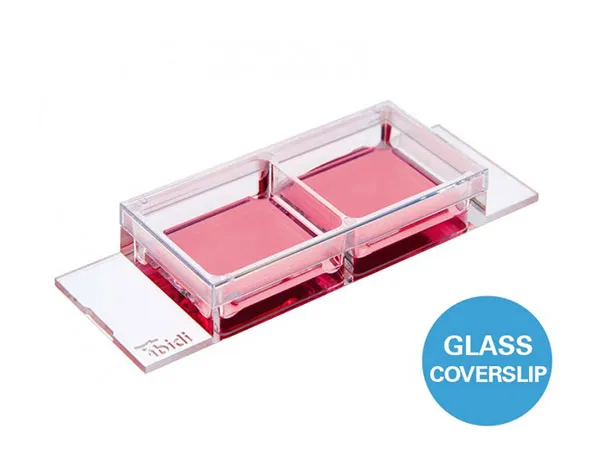
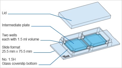
Chamber slide แบบ 2 ช่อง ก้นแก้ว – ประกอบด้วย intermediate plate เหมาะสำหรับเทคนิคแสงแบบ Phase contrast และกล้องจุลทรรศน์ชนิด TIRF
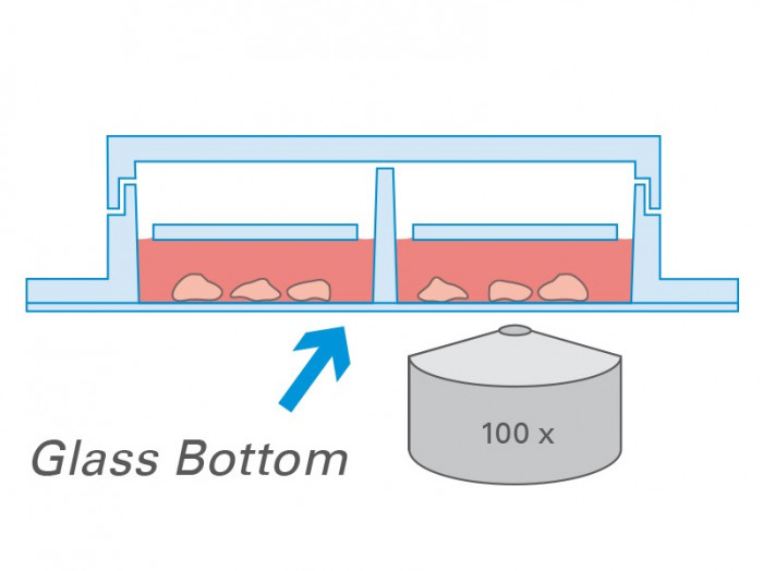
- การถ่ายภาพที่ยอดเยี่ยมเนื่องจากก้นภาชนะทำมาจากแก้วที่มีความบางเท่ากับ coverslip
- ประหยัดค่าใช้จ่ายในการทำการทดลองเนื่องจาก สามารถใช้เซลล์จำนวนน้อย และ reagent ปริมาณน้อย
- เหมาะสำหรับ Phase contrast โดยไม่ทำให้เกิด meniscus
A chambered coverslip with 2 wells, a #1.5H glass bottom, and a special intermediate plate for excellent phase contrast, suitable for use in TIRF and single molecule applications
- Perfect cell imaging thanks to the low thickness variability of the coverslips’ glass
- Cost-effective experiments using small numbers of cells and low volumes of reagents
- Excellent phase contrast over the entire well – no meniscus
- Surface Modification: #1.5H (170 µm +/- 5 µm) D 263 M Schott glass, sterilized
Pcs./Box: 15 (individually packed)
Applications
- Cultivation and microscopy of cell cultures
- TIRF and single molecule applications
Specifications
| Outer dimensions in mm (w x l) | 25.5 x 75.5 |
| Number of wells | 2 |
| Dimensions of wells (w x l x h) in mm | 21.6 x 23.8 x 9.3 |
| Volume per well | 1500 µl |
| Liquid height | 3.0 mm |
| Total height with / without lid | 10.8 / 9.5 mm |
| Growth area per well | 5.1 cm² |
| Coating area per well | 11.4 cm² |
| Bottom: Glass coverslip No. 1.5H, selected quality, 170 µm +/- 5 µm | |
Technical Features
- Open µ-Slide with 2 independent wells
- Intermediate plate in each well to avoid meniscus formation
- Ph+ feature for perfect phase contrast—no meniscus formation
- Bottom made from D 263 M Schott glass, No. 1.5H (170 +/- 5 µm)
- May require coating to promote cell attachment
- Also available as a µ-Slide 2 Well Ph+ with an ibidi Polymer Coverslip Bottom for superior cell growth
Experimental Example
ibidi Polymer Coverslip vs. ibidi Glass Bottom
ibidi Polymer Coverslip | ibidi Glass Bottom | |
| Optical properties | ||
| Refractive index (nD 589 nm) | 1.52 | 1.52 |
| Abbe number | 56 | 55 |
| Thickness | #1.5 (180 µm) | #1.5H (170 µm) |
| Material | Microscopy plastic | D 263M Schott borosilicate glass |
| Autofluorescence | Low | Low |
| Transmission | Very high (even ultraviolet) | High (ultraviolet restrictions) |
| Birefringence (DIC) | Low (DIC compatible*) | Low (DIC compatible*) |
| Other aspects | ||
| Surface modifications | ibiTreat – tissue culture treated Uncoated – hydrophobic | Only glass |
| Protein coatings | Compatible | Compatible |
| Gas permeable | Yes | No |
| Material flexibility | High | Low |
| Breakable | No | Yes |
| Applications | Fluorescence microscopy | TIRF and single photon |
Phase Contrast Microscopy
Phase contrast microscopy is the most commonly used, transmitted light technique in cell culture. When working with phase contrast microscopy, it is crucial to have the two phase rings adjusted to each other. In open wells, the meniscus at the air-water-interphase works like a lens that refracts the beam path. This miscalibrates the phase rings, leading to poor contrast in the microscopic image.
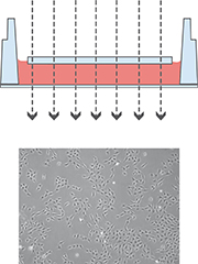
Working with the µ-Slide 2 Well Ph+ diminishes the meniscus, so that the whole optical system is aligned, no matter which position of the well is being imaged.
| µ-Slide 2 Well Glass Bottom  | µ-Slide 2 Well Ph+ Glass Bottom  |
Top View |  |
The µ-Slide 2 Well Ph+ Blass Bottom (Phase Contrast +) is designed for excellent optical quality, especially for cell culture when normal phase contrast microscopy is being used. Opposed to the classic µ-Slide 2 Well Glass Bottom, the Ph+ version provides a special intermediate plate in the center of the well. This plate flattens the meniscus that disturbs the phase contrast effect in normal open wells. Openings near the corners provide access to the wells for easy filling and aspiration of liquids.
This innovative technique supports meniscus-free phase contrast microscopy in a very convenient manner.
Working with the µ-Slide 2 Well Ph+ Glass Bottom diminishes the meniscus and increases the area of well contrasted cells
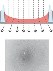 |  |
The illustration on the left shows the perturbing effect of a meniscus. Light is refracted on the air-water-interface, leading to poor contrast in microscopy. Only the small center part exhibits satisfying phase contrast.
Working with the µ-Slide 2 Well Ph+ Glass Bottom diminishes the meniscus and increases the area of well contrasted cells. The good contrast is due to the parallel beam path that is created by the plate.
