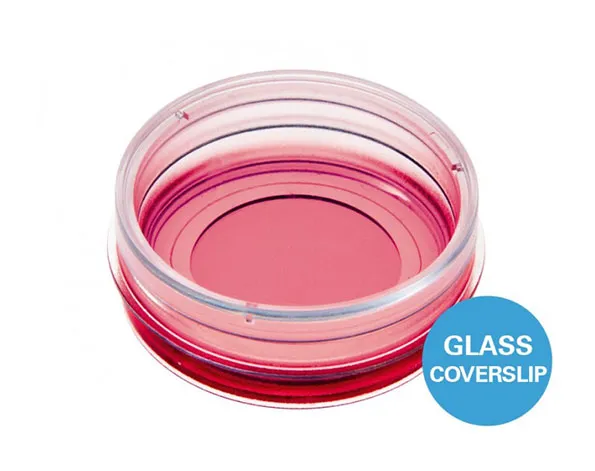
จานเพาะเลี้ยงเซลล์แบบก้นแก้ว สำหรับการถ่ายภาพ ขนาด 35 มม. – เหมาะสำหรับกล้องจุลทรรศน์ชนิด TIRF
- การถ่ายภาพที่ยอดเยี่ยมเนื่องจากก้นภาชนะทำมาจากแก้วที่มีความบางเท่ากับ coverslip (# 1.5H)
- มีฝาปิด พร้อมตำแหน่งล็อคที่ขอบจาน ช่วยลดการระเหย
- เหมาะสำหรับเทคนิค DIC เมื่อใช้ร่วมกับฝาปิดพิเศษสำหรับเทคนิค DIC
- นอกจากนี้ยังมีแบบมี grid 50 ไมครอน และ 500 ไมครอน
A 35 mm imaging dish with a glass bottom for use in TIRF, single molecule and super-resolution microscopy applications
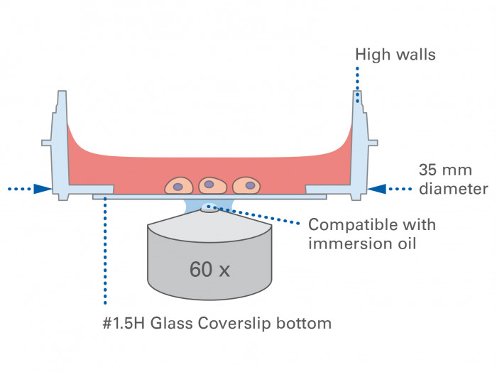
- In this microscopy dish, the cells are imaged on a high-quality No. 1.5H Glass Coverslip Bottom with very low thickness variability
- Cell culture dish equipped with a tightly fitting lid that minimizes evaporation
- Suitable for DIC when using a DIC Lid
- Surface Modification: #1.5H (170 µm +/- 5 µm) D 263 M Schott glass, sterilized
Pcs./Box: 60 (individually packed) - Surface Modification: #1.5H (170 µm +/- 5 µm) D 263 M Schott glass, sterilized
Pcs./Box: 400 (individually packed)
Applications
- Cultivation and high-resolution microscopy of cells
- Total Interference Reflection Fluorescence (TIRF) and single molecule applications
- Super-Resolution Microscopy (STED, SIM, (F)PALM, (d)STORM) and Fluorescence Correlation Spectroscopy (FCS)
- Immunofluorescence staining
- Widefield and confocal fluorescence microscopy of living and fixed cells
- Live cell imaging over extended time periods
- Transfection assays
- Differential interference contrast (DIC) when using a DIC Lid
- Cell location and counting when using the gridded version
Want to know if you should use a glass or a polymer bottom for your application? Find out here.
Specifications
| Ø µ-Dish | 35 mm |
| Volume | 2 ml |
| Growth area | 3.5 cm2 |
| Coating area using 400 µl | 4.1 cm2 |
| Ø observation area | 21 mm |
| Height with / without lid | 14/12 mm |
| Bottom: Glass Coverslip No. 1.5H, selected quality, 170 µm +/- 5 µm | |
Technical Features
- Standard format imaging dish with a 35 mm diameter for tissue culture
- Bottom made from D 263 M Schott glass with a thickness of 170 µm +/- 5 µm
- May require coating to promote cell attachment
- High walls with a standard height for easy handling
- Lid with locking feature for minimal evaporation
- Rim for easy opening
- No autofluorescence
- Fully biocompatible materials
- Available as a Bulk Pack with 400 individually packed µ-Dishes per box
- Available with a #1.5 ibidi Polymer Coverslip Bottom with optimized adhesion: µ-Dish 35 mm, high, for everyday use with diverse application possibilities
- Available with a 50 and 500 µm grid for cell counting: µ-Dish 35 mm, high Grid-50 Glass Bottom and µ-Dish 35 mm, high Grid-500 Glass Bottom
Test our everyday solution for cost-effective high-throughput experiments: Glass Bottom Dish 35 mm
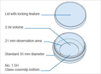

The Principle of the µ-Dish 35 mm, high Glass Bottom
The Coverslip Bottom
The µ-Dish 35 mm, high Glass Bottom comes with a thin #1.5H Glass Coverslip Bottom made from D 263 M Schott borosilicate glass that has the highest optical quality. ibidi developed these glass surfaces specifically for TIRF, super-resolution microscopy, and single molecule microscopy. The dish is also available with an #1.5 ibidi Polymer Coverslip Bottom for optimal cell adhesion and high-resolution microscopy in a variety of assays.
As another option for cost-effective high-throughput experiments, we offer the Glass Bottom Dish 35 mm with a #1.5 glass coverslip bottom.
Learn more about the coverslip bottom or the different surfaces of the ibidi chambers.
Find a comparison of the different ibidi Dishes 35 mm here.
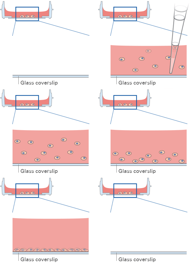
Lid with Locking Feature for Minimized Evaporation
All ibidi µ-Dishes are equipped with the special lid-locking feature. The locking position minimizes evaporation, and thereby provides excellent conditions for long-term studies in a non-humidified environment. Gas exchange (carbon dioxide or oxygen) during cell culture is maintained thanks to the gas-permeable plastic material of the dish.
TIP: Use the locking feature if minimal evaporation is required (e.g., outside incubators, non-humidified microscopy stages, etc.).

Experimental Examples

Time lapse of a B16-F1 mouse melanoma cell migrating on the laminin-coated surface of the µ-Dish 35 mm, high Glass Bottom. Phase contrast microscopy images were acquired over a a period of 10 minutes in the ibidi Stage Top Incubator Slide/Dish, CO2 – Silver Line using a 100x immersion objective on an Olympus X-81 microscope.
Data by Jonas Scholz and Prof. Jan Faix, Hannover Medical School, Germany.
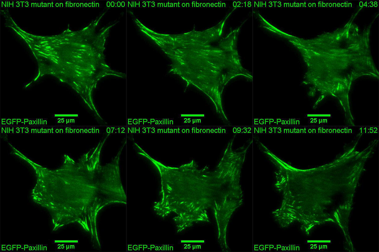
Time lapse TIRF imaging showing focal adhesion dynamics of a mutant NIH-3T3 mouse embryonic fibroblast cell, which was transfected with EGFP-Paxillin, in a µ-Dish 35 mm, high Glass Bottom. TIRF microscopy images were acquired over a period of 12 hours in the ibidi Stage Top Incubator Slide/Dish, CO2 – Silver Line using a TIRF 100x immersion objective on a Nikon Eclipse microscope.
Data by Jonas Scholz and Prof. Jan Faix, Hannover Medical School, Germany.
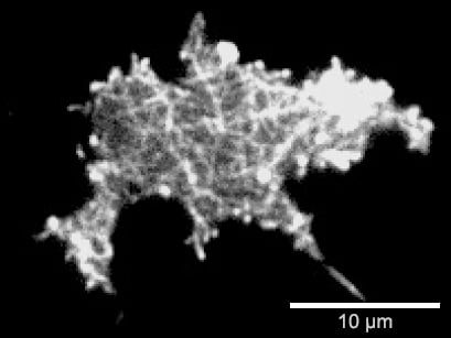
Surface-near F-actin network of a Dictyostelium discoideum DdLimE-GFP cell. Live cell imaging on a Glass Coverslip #1.5H using Total Interference Reflection Fluorescence (TIRF) microscopy.

Fluorescence microscopy of fixed rat fibroblasts on a Glass Coverslip #1.5H. F-actin filaments are stained with phalloidin (green), nuclei are stained with DAPI (blue).
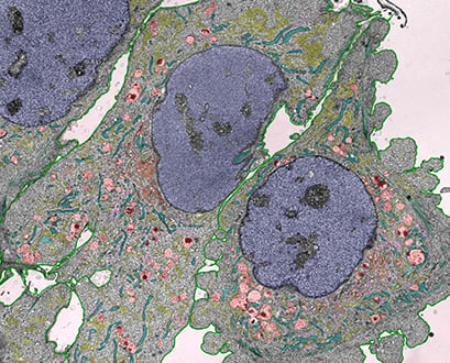
Electron microscopy of longitudinal ultra-thin sections of mouse embryonic fibroblasts (MEFs). Before fixation and processing, the cells were cultivated on an ibidi μ-Dish 35 mm, high Glass Bottom. STEM imaging was done using a FEI Verios 460L. Structures are indicated by color overlays: lysosomes (red), mitochondria (cyan), ER (yellow), golgi (orange), microtubules (magenta), plasma membrane (green), nucleus (blue), actin (turquoise). Image by Christian Lamberz, University of Bonn, German Centre for Neurodegenerative Diseases (DZNE), Germany.
Comparison of the ibidi Dishes 35 mm
Please find a detailed comparison of the material specifications, including suitable microscopy applications for the ibidi Polymer Coverslip, the ibidi Glass Bottom, and further materials here.
| Bottom material | #1.5 ibidi Polymer Coverslip | #1.5H ibidi Glass Coverslip | #1.5 glass coverslip |
| Bottom thickness | 180 µm (+10/-5 µm) | 170 µm (+/-5 µm) | 170 µm (+20/-10 µm) |
| Bottom: gas permeability | Yes | No | No |
| Available surfaces | ibiTreat (tissue culture treated), Uncoated (hydrophobic) | Uncoated glass | Uncoated glass |
| Lid | Lid with locking feature | Lid with locking feature | Standard lid |
| Packaging | Sterile, individually packed | Sterile, individually packed | Sterile, 10 pcs. per case |
| Quantity | 60 or 400 pcs./box | 60 or 400 pcs./box | 200 or 800 pcs./box |
| Low wall version available? | Yes: µ-Dish 35 mm, low | Yes: µ-Dish 35 mm, low Glass Bottom | No |
| Gridded version available? | Yes: µ-Dish 35 mm, high Grid-500 | Yes: µ-Dish 35 mm, high Grid-50 Glass Bottom, µ-Dish 35 mm, high Grid-500 Glass Bottom | No |
