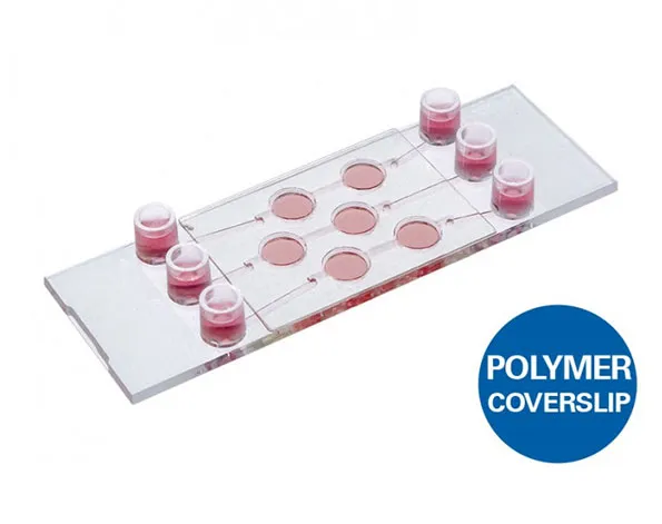
µ-Slide แบบ 3 ช่องสำหรับการเพาะเลี้ยงเซลล์ หรือการกระจายของเซลล์ที่เป็น 3 มิติ
- เหมาะสำหรับการเพาะเลี้ยงเซลล์แบบ 3 มิติในระยะยาว
- ใช้งานง่าย ตัวอย่างสามารถใส่ในหลุมที่เปิดอยู่ และการกระจายตัวจะเกิดขึ้นหลังจากการปิดหลุม
- ภาพที่ได้มีคุณภาพสูง สามารถถ่ายภาพความละเอียดสูงได้
A three channel µ-Slide used for the cultivation and perfusion of 3D cell structures
- Ideal for the long-term cultivation of cells in 3D matrices with an optimal nutrients supply
- Easy handling: Samples can be placed into the open wells–perfusion can be applied after closing the wells
- Excellent optical-quality imaging chamber for high-end microscopy
- Surface Modification: Uncoated: #1.5 polymer coverslip, hydrophobic, sterilized
Pcs./Box: 15 (individually packed) - Surface Modification: ibiTreat: #1.5 polymer coverslip, tissue culture treated, sterilized
Pcs./Box: 15 (individually packed)
Applications
- Observation of single cells in 3D matrices or tissue samples (e.g., spheroids, small organoids, or organisms)
- Perfusion of samples
- Long-term cultivation of cells in 3D matrices
Specifications
| Outer dimensions (w x l) | 25.5 x 75.5 mm² |
| Number of wells | 6 |
| Volume of wells | 30 µl |
| Well diameter | 5.5 mm |
| Well height (without channel) | 1.2 mm |
| Well height (with channel) | 1.7 mm |
| Growth area per well | 25 mm2 |
| Number of channels | 3 |
| Total channel volume | 130 µl |
| Channel width | 1.0 mm |
| Adapters | Female Luer |
| Volume per reservoir | 60 µl |
| Coating area using 30 µl | 0.29 cm² per well |
| Coating area using 130 µl | 2.4 cm2 per channel |
| Top cover matches coverslip | No. 1.5 |
| Bottom | ibidi Polymer Coverslip |
Technical Features
- Open wells are filled and later closed for the perfusion of the sample
- Channels allow for easy fluid connection of the wells
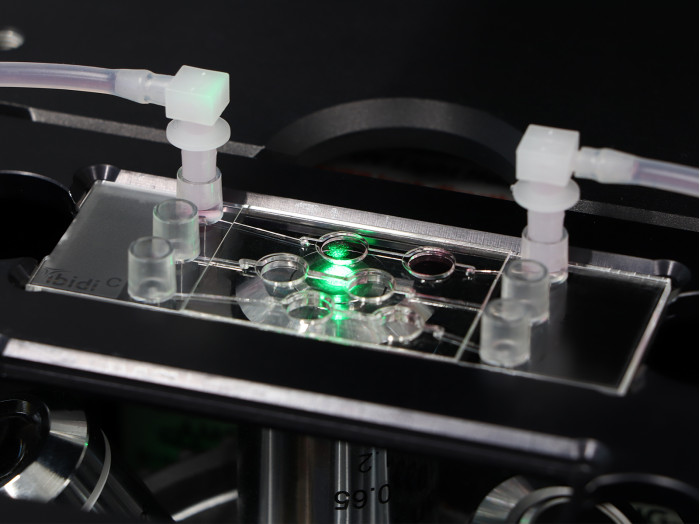
- Luer adapters for easy pump connection (e.g., to the ibidi Pump System)
- Optimal nutrients supply into 3D structures
- Observe the sample through the polymer coverslip bottom using high resolution light microscopy
The Principle of the µ-Slide 3D Perfusion
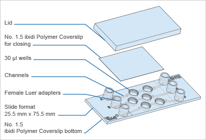
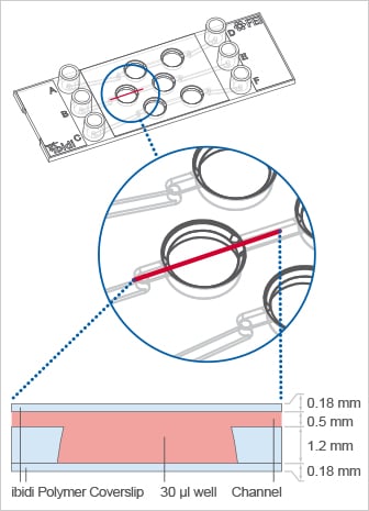
The µ-Slide 3D Perfusion contains six wells, in which cells can be cultivated in 3D matrices and imaged using high-resolution microscopy. Two of the six wells, respectively, are connected by a channel. For optimal nutrition of the cells during long-term culture, the channels can be connected to a pump for perfusing the wells.
Application Examples Using the µ-Slide 3D Perfusion
3D Matrix Culture
Single cells can be cultured and imaged in a 3D matrix, e.g., a collagen gel.

Soft Matrix Culture
Adherent cells can be cultured and imaged on a soft matrix.

Culture Without Matrix
Adherent cells can be seeded directly on the substrate without any matrix.

Spheroid Culture
Spheroids can be cultured and imaged, e.g., in a gel sandwich.

Tissue Sample Culture
Tissue samples can be cultured and imaged, e.g., in a gel sandwich.

Standard Filling Procedure

Which Slide Should I Use for My Application?
| µ-Slide Spheroid Perfusion | µ-Slide III 3D Perfusion | µ-Slide I Luer 3D | µ-Slide I Luer | |
| Application | ||||
| Perfusion of samples | Yes | Yes | Yes | Yes |
| Defined shear stress on cell monolayers | No | No | Yes, on gel matrix | Yes, on coverslip |
| Gel matrices for 3D | No | Yes | Yes | No |
| Cell Type | ||||
| Spheroids/organoids | Yes, free floating in well | Yes, inside gel matrix only | Yes, inside gel matrix only | No |
| Suspension cells | Yes, free floating in well | Yes, inside gel matrix only | Yes, inside gel matrix only | Yes, in flow suspension only |
| Adherent cells | Yes, on coverslip | Yes, inside or on gel matrix | Yes, inside or on gel matrix | Yes, on coverslip |
