ซิลิโคนแบบ 4 ช่อง เพื่อวางบนจานเลี้ยงเซลล์ – สำหรับการถ่ายภาพแบบ live cell ทั้งเซลล์เกาะติดและเซลล์แขวนลอย
- สำหรับการใช้กล้องจุลทรรศน์ในระยะยาว
- การเพาะเลี้ยงเซลล์จำนวนที่น้อยโดยปราศจากการระเหย
- ใน well มีลักษณะคล้ายรูปทรงกรวยเพื่อการมองภาพที่ยอดเยี่ยม
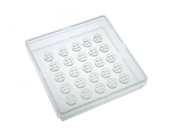
- มีซิลิโคนจำนวน 25 ชิ้น
- สำหรับวางลงบน 6 หรือ 12 well plates หรือขนาดอื่นๆ และสามารถวางบนจานเพาะเลี้ยงเซลล์
A 4 well silicone insert for live cell imaging of both adherent and suspension cells
- Allows long-term microscopy assays of single cells
- Cultivation of small numbers of cells without evaporation risks
- Conical wells for superb optical quality
- 25 pieces in a transport dish
- Used for self insertion into 6 or 12 well plates, or other formats
- Pcs./Box: 25 (in 1 case)
Product Variation: for self-insertion in a 10 cm transport dish
Please note: Not to be used without transferring the micro-Insert
Applications:
- Live cell imaging of adherent and suspension cells
- Immunofluorescence assays
- Immobilization of cells in small wells, e.g., for 3D assays
- Performing long-term microscopy assays lasting up to several weeks
- Co-cultivation of cells
- Cell tracking of very small cell numbers over time
- Cell differentiation, e.g., single stem cells
Specifications:
| Number of wells | 4 |
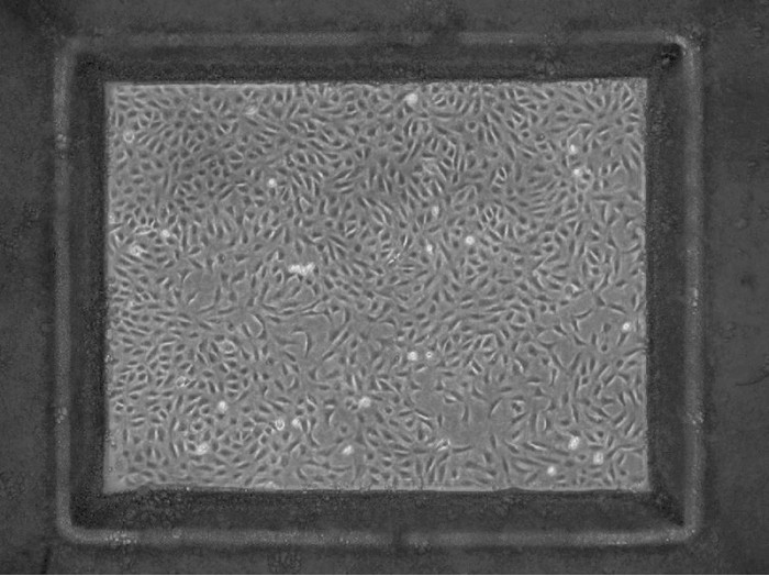
| Dimensions of wells (w x l) in mm | 2.0 x 1.5 (digital format 4:3) |
| Diameter of the complete insert | 12 mm |
| Height of the complete insert | 4.2 mm |
| Volume per well | 10 µl |
| Growth area per well | 0.03 cm2 |
| Coating area per well | 0.23 cm2 |
| Material | Biocompatible silicone |
| Bottom | No bottom – sticky underside |
Technical Features:
- Four small 2.0 mm x 1.5 mm microscopy wells with a 4:3 digital format
- Suspension cells cannot escape the observation field
- Adhesive underside
- Transferable to any flat clean surface
- No material is left behind upon the insert’s removal
- Includes a rim for easy handling of the insert with tweezers

Product Selection Guide
| micro-Insert 4 Well FulTrac 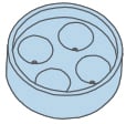 Single cells20x Objective lens for full well Single cells20x Objective lens for full wellcoverageHigh resolution fluorescence microscopy | micro-Insert 4 Well  Few cells4x Objective lens for full well Few cells4x Objective lens for full wellcoverageHigh resolution fluorescence microscopy |
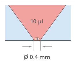 | 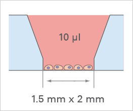 |
Cross Section

Ideal Coverage using Digital Imaging with CCD Cameras

3D Application:

Possible Filling Procedures:

Minor Well Filling
- Minimal volume
- Individual wells

Whole Insert Filling
- Moderate volume
- Wells connected by medium
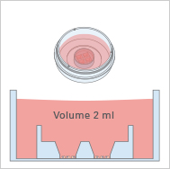
Whole µ-Dish Filling
- Maximum volume
- Maximum experimental duration

Evaporation Barrier
Co-Cultivation Assays

Coating patches (spots) with proteins

hydrophilic protein spots on hydrophobic, uncoated µ-Dish
