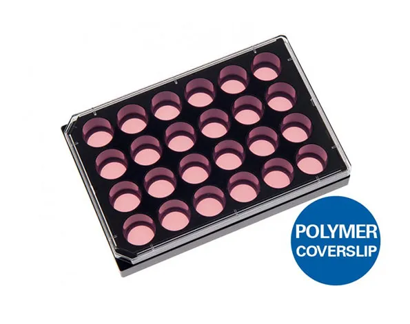
ไมโครเวลเพลทแบบก้นแบนและใสชนิด 24 หลุม สีดำ – สำหรับงานเพาะเลี้ยงเซลล์
- เหมาะสำหรับกล้องจุลทรรศน์
- ด้านในมีพื้นเรียบ
- ก่อให้เกิด crosstalk ต่ำในแต่ละ well ในการใช้กล้องจุลทรรศน์แบบฟลูออเรสเซนต์
A black 24 well plate with flat and clear bottom for high throughput microscopy
- Ideal for high resolution imaging through the No.1.5 polymer coverslip bottom with the highest optical quality
- Excellent inner well flatness and whole plate flatness
- Low well-to-well crosstalk in fluorescence microscopy
- Surface Modification: ibiTreat: #1.5 polymer coverslip, tissue culture treated, sterilized
Pcs./Box: 15 (individually packed) - Surface Modification: Uncoated: #1.5 polymer coverslip, hydrophobic, sterilized
Pcs./Box: 15 (individually packed)
Applications
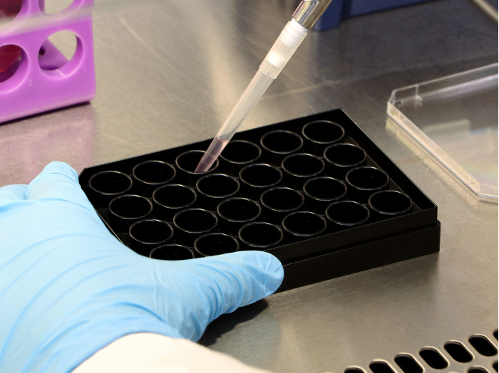
- Fluorescence-based imaging of living or fixed cells
- High throughput screening (HTS) in cell culture
- Wound healing experiments with ibidi Culture-Inserts
- Large-scale transfection experiments
Specifications
| Length / width | 127.5 / 85.5 mm |
| Height with / without lid | 22.4 / 20.0 mm |
| Well-to-well distance | 18.9 mm |
| Well clearance | 0.82 mm |
| Single well diameter | 14.0 mm |
| Single well depth | 19.0 mm |
| Volume per well | 1 ml |
| Growth area per well | 1.54 cm2 |
| Coating area per well using 1 ml | 4.4 cm2 |
| Bottom: ibidi Polymer Coverslip | |
Technical Features
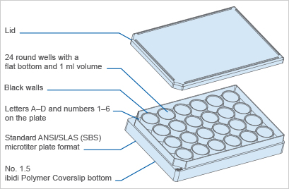
- Standard format, and dimensions of 85.5 x 127.5 mm (meets ANSI/SLAS (SBS) Standards)
- 24 round wells with standard numbering
- Compatible with automation equipment
- Excellent inner well flatness and whole plate flatness
- Suitable for fluorescence scanners
- High-quality plastic bottom, which is compatible with solvents for staining and fixation, and also with immersion oil
- Sterile, single packaging
Technical Drawing
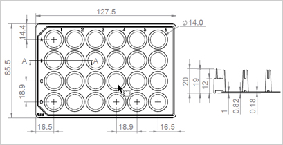
Technical drawings and details are available in the Instructions (PDF).
Experimental Examples
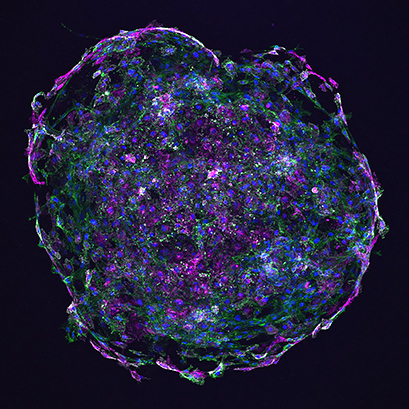
Bead sprouting assay shows irregular blood vessel formation of human umbilic venous endothelial cells (HUVECs). Cells were transduced with an mScarlet-tagged lentiviral plasmid (magenta), embedded into a fibrin gel in an ibidi µ-Plate 24 Well, and co-cultured with lung fibroblasts for 7 days. The actin cytoskeleton was stained with phalloidin (green) and nuclei were stained with DAPI (blue). The image was taken using a Zeiss LSM 710 Meta confocal scanner with a 10x objective. Image by Tish Essebier, Institute for Molecular Bioscience, The University of Queensland, Brisbane, Australia.

Confocal microscopy of rat hippocampal neurons plated over astrocytes, cultured on ibidi µ-Plate 24 Well (#82406). Red is Synapsin 1, a presynaptic marker, white is neuronspecific Tubulin Beta-III, and blue is DAPI, a nuclear counterstain. Image by Daniel Hoeppner, Lieber Institure for Brain Development, Baltimore, MD, USA.
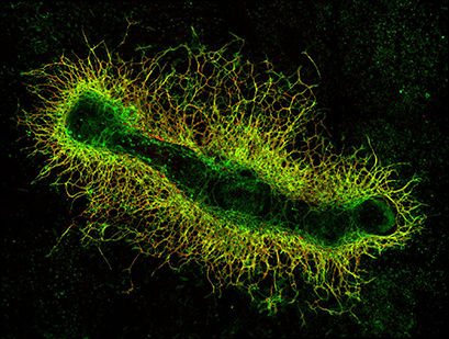
A murine metatarsal as an ex vivo model for sprouting angiogenesis. The fetal mouse bone was obtained from an E16.5 LifeAct-GFP mouse embryo and cultured for 14 days in an ibidi µ-Plate 24 Well, leading to the outgrowth of blood vessels. The metatarsal shows the actin cytoskeleton in green and was stained for CD31 (PECAM-1) in red as an endothelial marker to highlight vessel outgrowth from the bone. Image by Lilian Schimmel and Emma Gordon, Institute for Molecular Bioscience, The University of Queensland, Brisbane, Australia.
