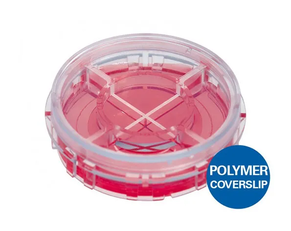
จานเพาะเลี้ยงเซลล์แบบแบ่ง 4 ช่อง ก้นบางแบบพอลิเมอร์พิเศษ ขนาด 35 มม. – เหมาะสำหรับการถ่ายภาพด้วยกล้องจุลทรรศน์
- จานเพาะเลี้ยงเซลล์แบ่งออกเป็น 4 ช่อง สำหรับทำการทดลองแยกกัน
- เหมาะสำหรับเทคนิคแสงแบบเฟสคอนทราสต์
- มีเทคโนโลยี ibiTreat surface เพื่อการเจริญเติบโตของเซลล์ในอุดมคติ
- ก้นภาชนะทำมาจากพอลิเมอร์พิเศษของ ibidi มีความบางเท่ากับ coverslip
A 35 mm imaging dish with four compartments and the ibidi Polymer Coverslip Bottom—for simultaneous cellular assays and high end microscopy
- Perform up to four simultaneous, individual experiments in the subdivisions of the dish
- Superior phase contrast and homogeneous cell growth due to the Ph+ feature
- Unique ibidi Polymer Coverslip Bottom that combines both:
– Excellent cell culture conditions
– Supreme optical quality for high resolution microscopy - Ideal cell growth conditions provided by the ibiTreat surface
- Surface Modification: Uncoated: #1.5 polymer coverslip, hydrophobic, sterilized
Pcs./Box: 60 (individually packed) - Surface Modification: ibiTreat: #1.5 polymer coverslip, tissue culture treated, sterilized
Pcs./Box: 60 (individually packed)
Applications
- Cell culture and high resolution fluorescence microscopy
- Simultaneous multiplex analysis of, for example, different cell lines or distinct experimental conditions
- Immunofluorescence staining
- Live cell imaging
- Transfection
Specifications
| Ø µ-Dish | 35 mm |
| Volume per well | 300 µl |
| Liquid height | 4.0 mm |
| Growth area per well | 0.85 cm² |
| Coating area per well | 2.46 cm² |
| Partition wall | 0.6 mm |
| Diameter growth area | 21 mm |
| Height with/without lid | 12/10 mm |
| Bottom: ibidi Polymer Coverslip |
Technical Features
- Subdivided version of the µ-Dish 35 mm, high
- Four separated wells in one petri dish
- Marked compartments for easy orientation
- Small area for minimization of required reagents and cells
- Ph+ feature for perfect phase contrast—no meniscus formation
- Made of biocompatible material without glues
- Integrated rim for easy opening
- No autofluorescence
- Compatible with all staining and fixation solvents
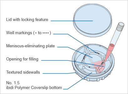
Subdivided Wells for 4 Individual Experiments in One Dish
Being divided into 4 chambers, the µ-Dish 35mm Quad quadruples the sample number and throughput. This feature makes it ideal for simultaneous analysis of individual cell lines or different experimental conditions—all on one single plate. In addition to supporting applications such as immunofluorescent staining and transfection, which can be performed in standard 4 chamber dishes, ibidi’s quadrant petri dish also offers highly superior optics.
Centered Plate for Superior Phase Contrast
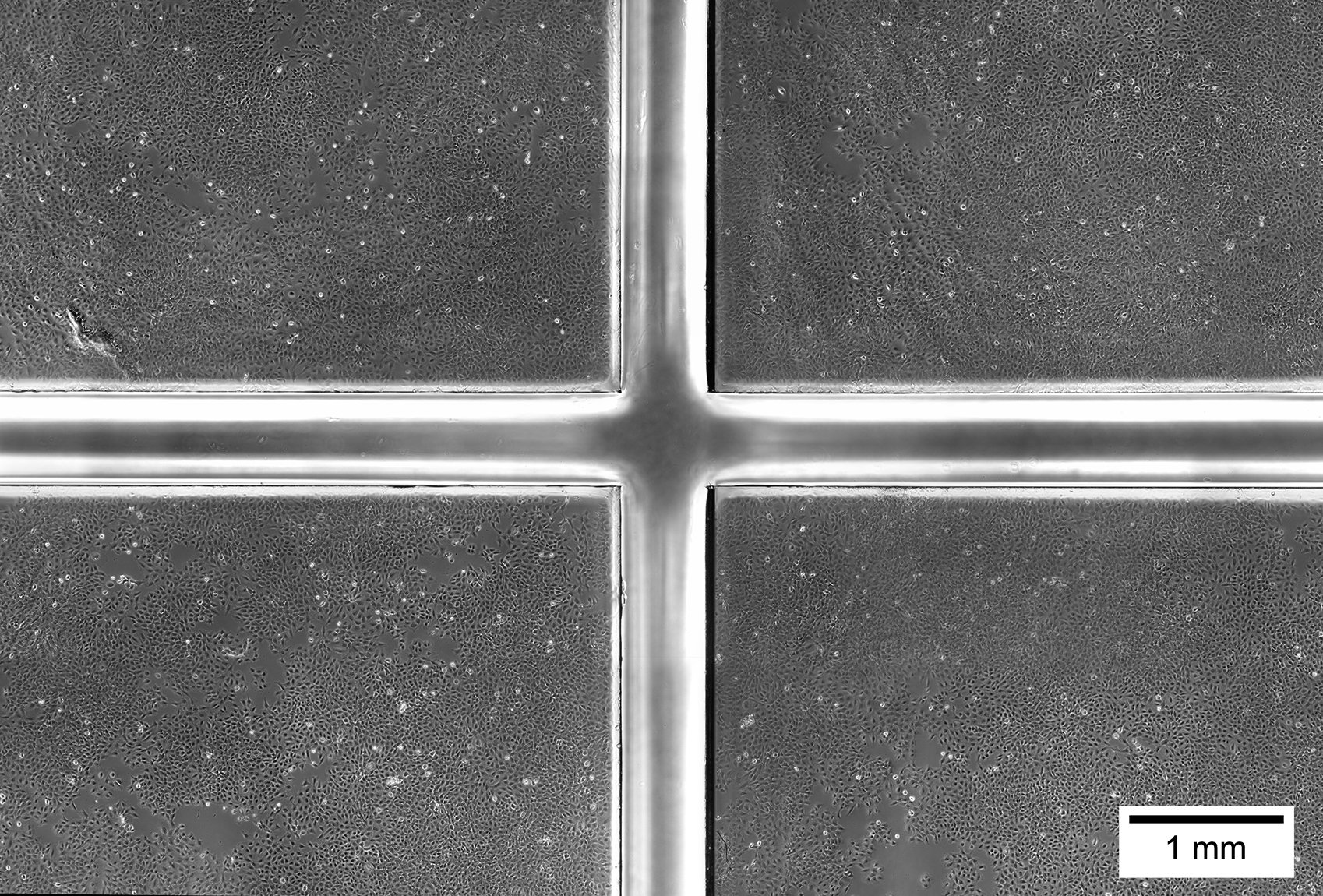
The centered, intermediate plate diminishes meniscus formation, which enables brilliant phase contrast optics. At the same time, this Ph+ feature facilitates homogeneous cell distribution.
Just like the ibidi µ-Slide 2 Well Ph+ and the µ-Slide 4 Well Ph+, the µ-Dish 35mm Quad combines excellent cell growth and brilliant high resolution microscopy—no matter if phase contrast or fluorescence microscopy is used.
Small Imaging Area for Minimized Stage Travelling
The ibidi µ-Dish 35mm Quad guarantees minimized travel ranges of microscope stages and objective lenses. Due to the thin partition wall that separates the different wells, the cells in each well can be rapidly imaged with a minimum of stage/objective travelling. The ibidi µ-Dish 35mm Quad was invented by ibidi and introduced into the market by Nikon in 2007 as Hi-Q4 culture dish. It has already proven its suitability on the Nikon BioStation IM and BioStation IM-Q, which are widely used systems for live cell screening.
µ-Dish 35 mm Quad | µ-Dish 35 mm, high |
Top View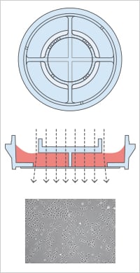 | . |
ibidi Polymer Coverslip Bottom for Brilliant Optics
Being divided into 4 chambers, the µ-Dish 35mm Quad quadruples the sample number and throughput. This feature makes it ideal for simultaneous analysis of individual cell lines or different experimental conditions—all on one single plate. In addition to supporting applications such as immunofluorescent staining and transfection, which can be performed in standard 4 chamber dishes, the ibidi quadrant petri dish also offers highly superior optics.
ibiTreat Surface for Optimal Cell Adherence
µ-Dishes, with the tissue culture-treated, hydrophilic ibiTreat surface, are perfectly suited for the culture and microscopy of many adherent cell types. The unique ibiTreat surface provides ideal cell growth conditions for the most adherent cell types, without further coatings.

Triple immunofluorescence of MDCK cells
Red: mitochondria, stained with MitoTracker™ Red CMXRos
Green: F-actin, stained with Alexa Fluor™ 488 Phalloidin
Blue: nuclei, stained with DAPI
Lid with Locking Feature for Minimized Evaporation
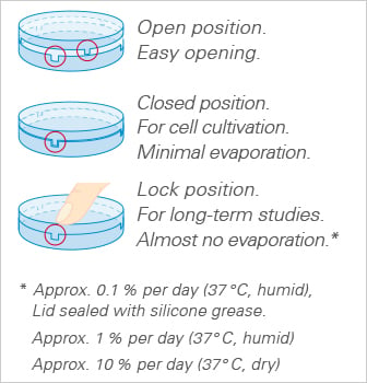
All ibidi µ-Dishes are equipped with the special lid locking feature. The locking position minimizes evaporation and thereby provides excellent conditions for long-term studies in a non-humidified environment. Gas exchange (carbon dioxide, or oxygen) during cell culture is maintained thanks to the gas-permeable plastic material of the dish.
TIP: Use the locking feature only if minimal evaporation is required (e.g., outside incubators, non-humidified microscopy stages, etc.).
Excellent Phase Contrast in the µ-Dish 35 mm Quad
